
















©The Author(s) 2020.
World J Clin Cases. Jan 26, 2020; 8(2): 362-369
Published online Jan 26, 2020. doi: 10.12998/wjcc.v8.i2.362
Published online Jan 26, 2020. doi: 10.12998/wjcc.v8.i2.362
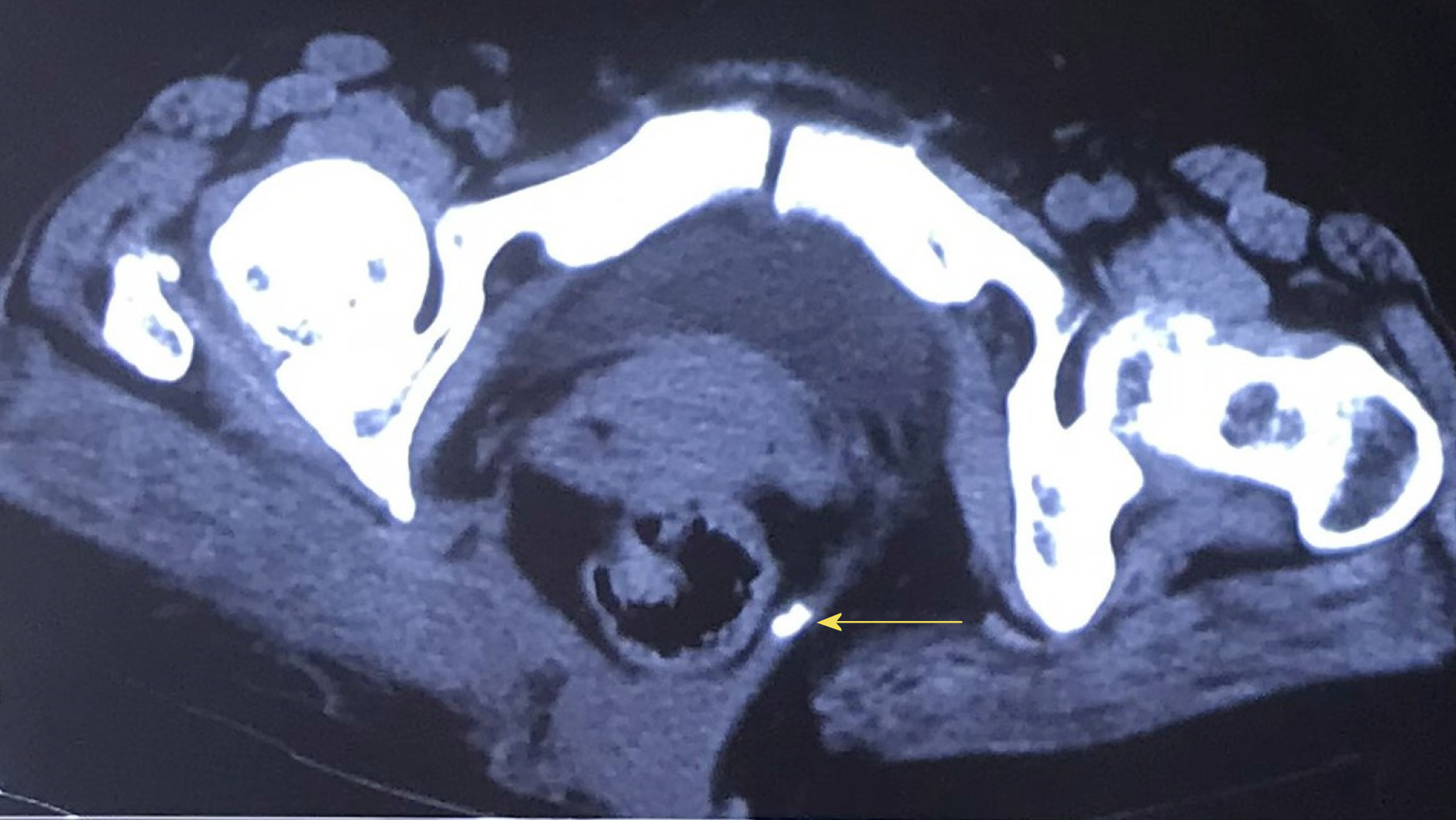
Figure 2 Computed tomography image.
Computed tomography revealed rectal herniation and an abnormality in the structure of the tissues between the sacrococcyx (arrow) and the right gluteus muscle.
- Citation: Dong YQ, Liu LJ, Fu Z, Chen SM. Mesh repair of sacrococcygeal hernia via a combined laparoscopic and sacrococcygeal approach: A case report. World J Clin Cases 2020; 8(2): 362-369
- URL: https://www.wjgnet.com/2307-8960/full/v8/i2/362.htm
- DOI: https://dx.doi.org/10.12998/wjcc.v8.i2.362