Copyright
©The Author(s) 2020.
World J Clin Cases. Jan 26, 2020; 8(2): 318-324
Published online Jan 26, 2020. doi: 10.12998/wjcc.v8.i2.318
Published online Jan 26, 2020. doi: 10.12998/wjcc.v8.i2.318
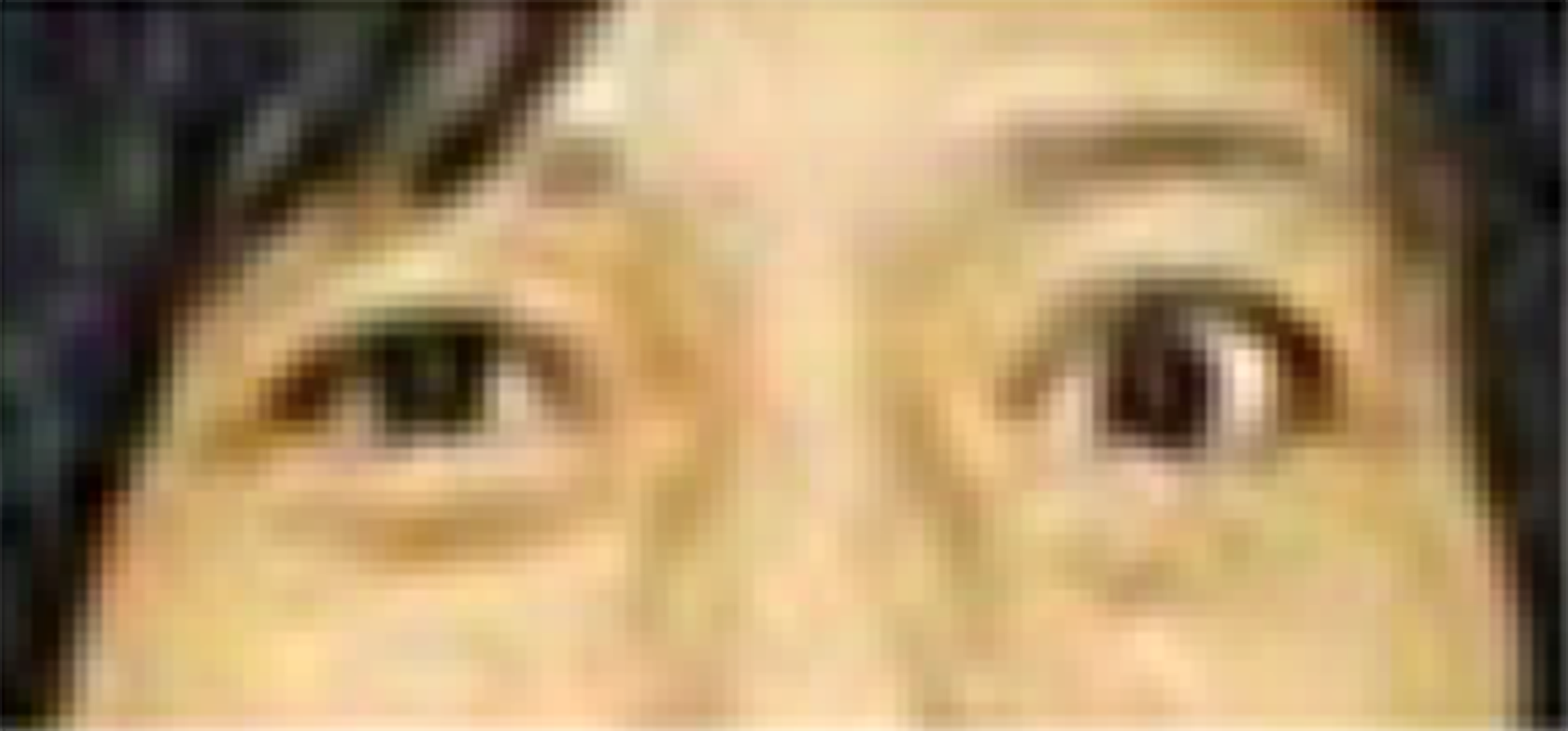
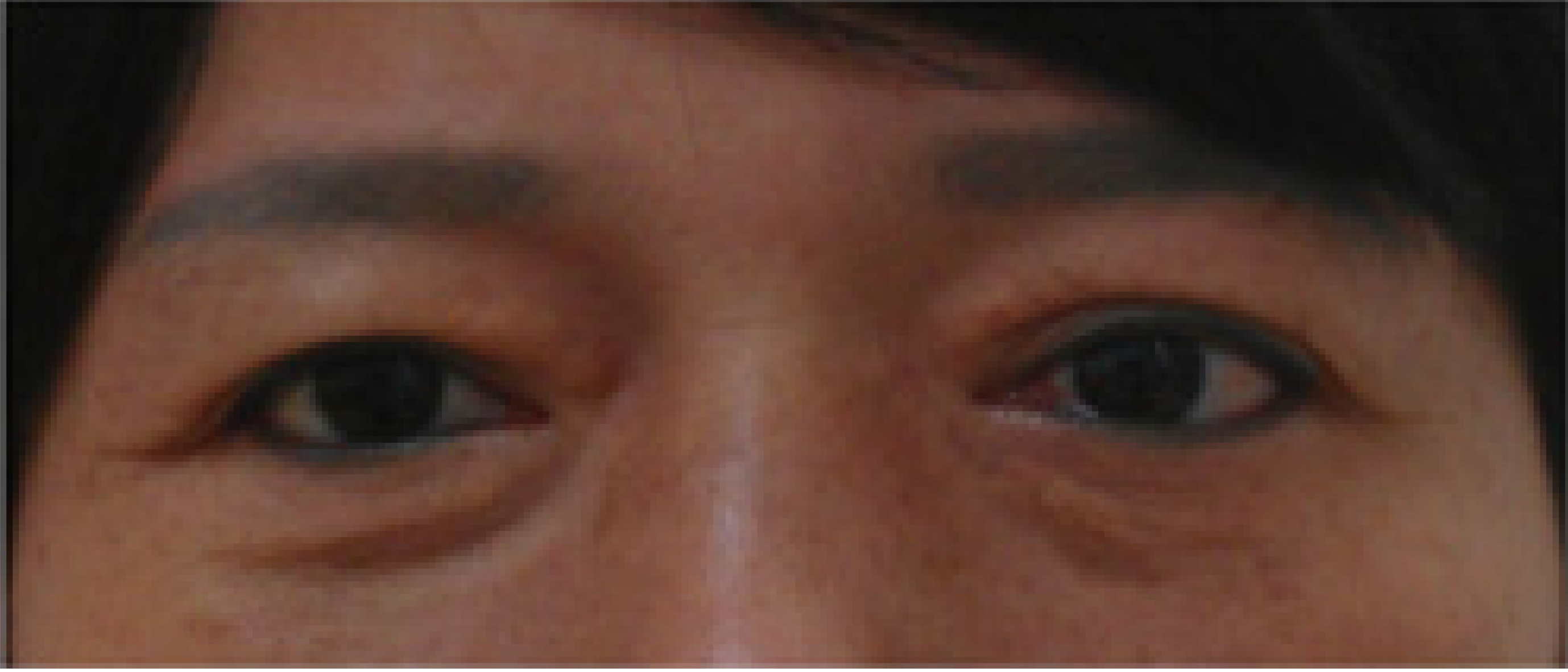
Figure 1 The pre-operative photograph revealed unilateral exophthalmos over the left eye.
The distance from upper to lower marginal reflex of left eye was 13 mm (vs 10 mm for the right eye).
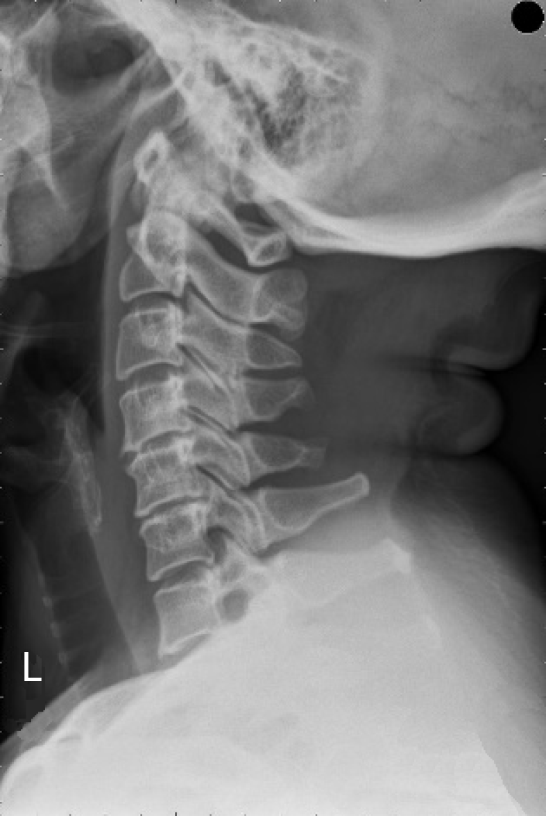
Figure 2 X-ray revealed marginal osteophytes with disc space narrowing at C4-5-6 vertebrae.
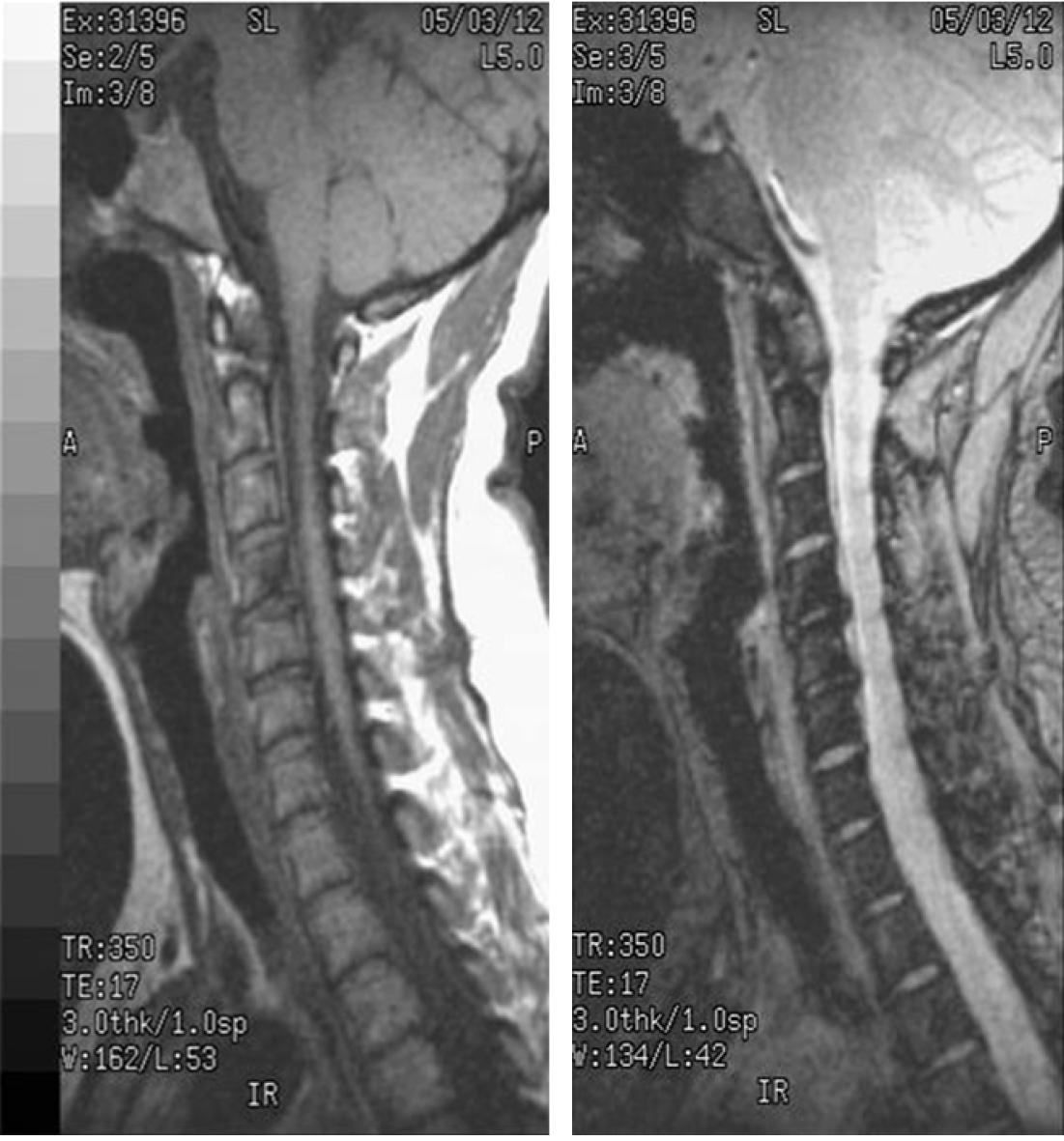
Figure 3 Magnetic resonance imaging showed posterior disc protrusion causing cervical cord compression at C4-5-6 vertebrae.
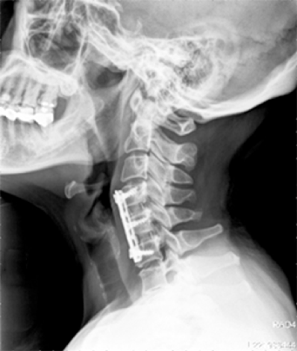
Figure 4 The X-ray showed that the patient underwent an operation involving an anterior cervical diskectomy and fixation with two PEEK interbody cages and a plate at C4/5/6 vertebrae.
Figure 5 The post-operative picture illustrated left unilateral exophthalmos improvement spontaneously.
The distance from upper to lower marginal reflex of left eye was 10 mm.
- Citation: Wu CM, Liao HE, Hsu SW, Lan SJ. Cervicogenic exophthalmos: Possible etiology and pathogenesis. World J Clin Cases 2020; 8(2): 318-324
- URL: https://www.wjgnet.com/2307-8960/full/v8/i2/318.htm
- DOI: https://dx.doi.org/10.12998/wjcc.v8.i2.318


