Copyright
©The Author(s) 2020.
World J Clin Cases. Oct 6, 2020; 8(19): 4676-4680
Published online Oct 6, 2020. doi: 10.12998/wjcc.v8.i19.4676
Published online Oct 6, 2020. doi: 10.12998/wjcc.v8.i19.4676
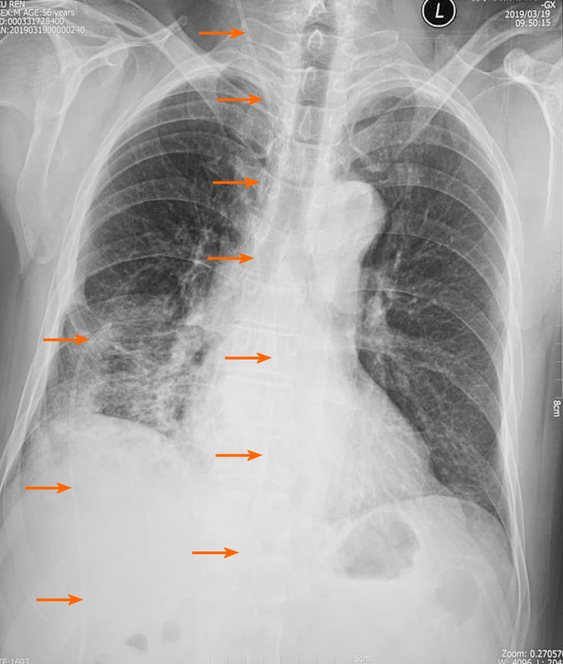
Figure 1 X-ray examination revealed that the peritoneal end of the shunt tube moved to the pleural cavity with the formation of pulmonary inflammation on March 25, 2019 (The orange arrows indicate the shunt tube).
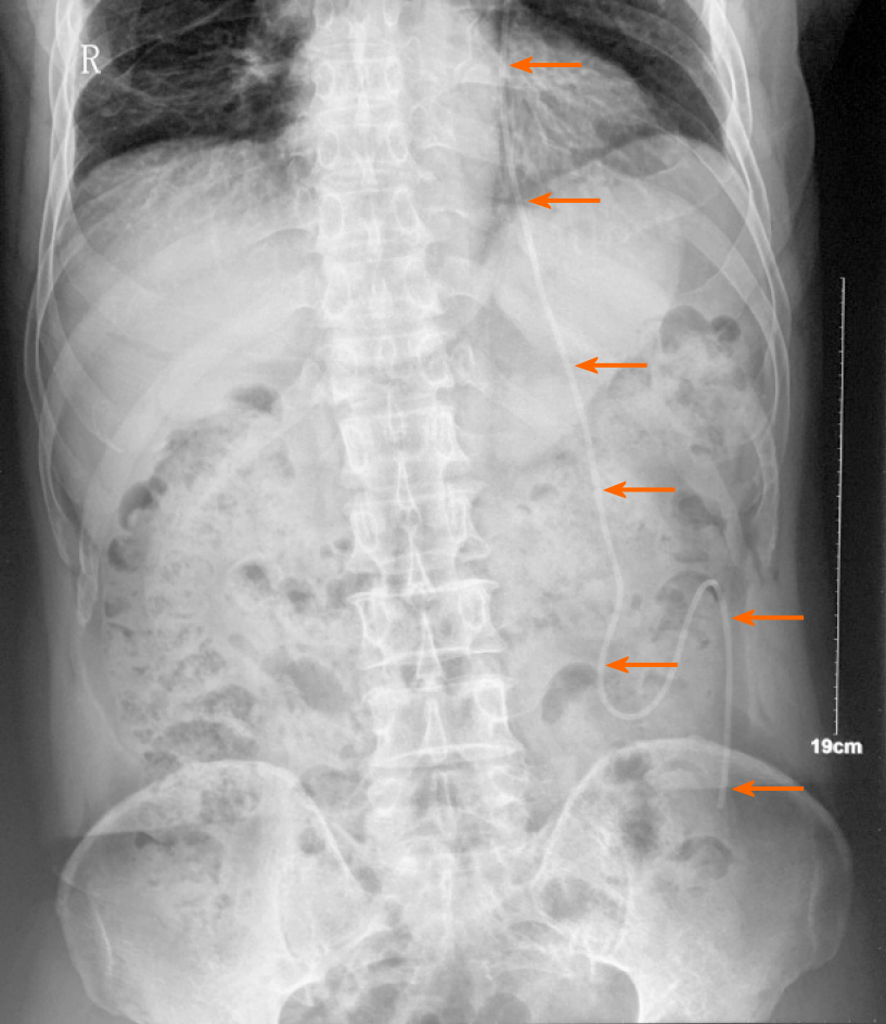
Figure 2 X-ray examination showed that the shunt tube was located in the abdominal cavity after operation on April 4 (The orange arrows indicate the shunt tube).
- Citation: Liu J, Guo M. Displacement of peritoneal end of a shunt tube to pleural cavity: A case report. World J Clin Cases 2020; 8(19): 4676-4680
- URL: https://www.wjgnet.com/2307-8960/full/v8/i19/4676.htm
- DOI: https://dx.doi.org/10.12998/wjcc.v8.i19.4676


