Copyright
©The Author(s) 2020.
World J Clin Cases. Oct 6, 2020; 8(19): 4544-4549
Published online Oct 6, 2020. doi: 10.12998/wjcc.v8.i19.4544
Published online Oct 6, 2020. doi: 10.12998/wjcc.v8.i19.4544
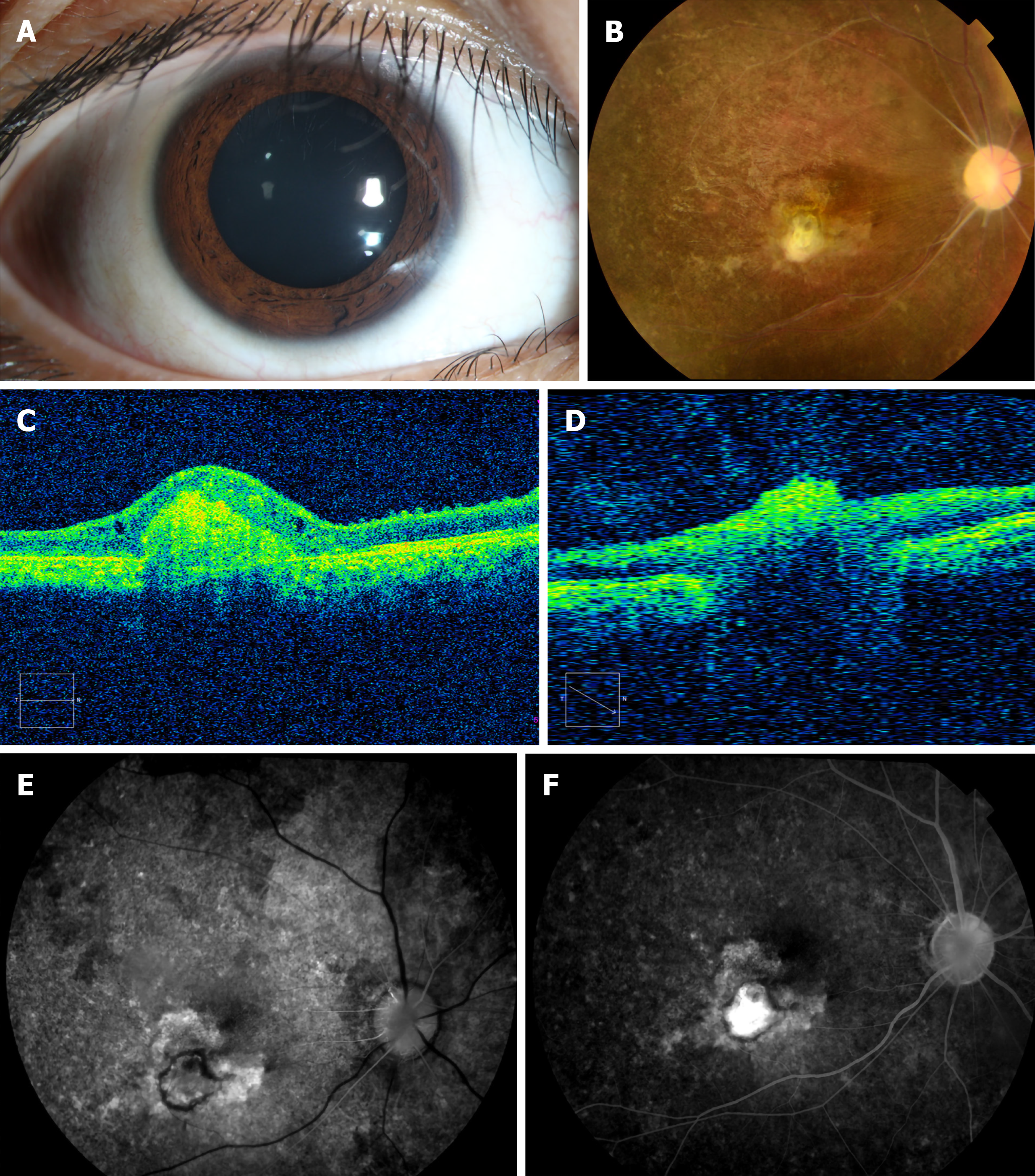
Figure 1 The boy’s ocular examination results at presentation.
A: The conjunctiva was 1+ injected in the right eye; B: Fundus images showed optic disc pallor, retinal vascular occlusion, pigmentation of the retina and a gray-white lesion (scar) in the right eye; C: Optical coherence tomography B-scans passing through the gray-white lesion showed an elevated lesion, and thinning of the whole retinal layer; D: Optical coherence tomography B-scans passing through the optic disk demonstrated a severely reduced peripapillary retinal nerve fiber layer; E: Fundus fluorescein angiography in the early phase; and F: Fundus fluorescein angiography in the late phase.
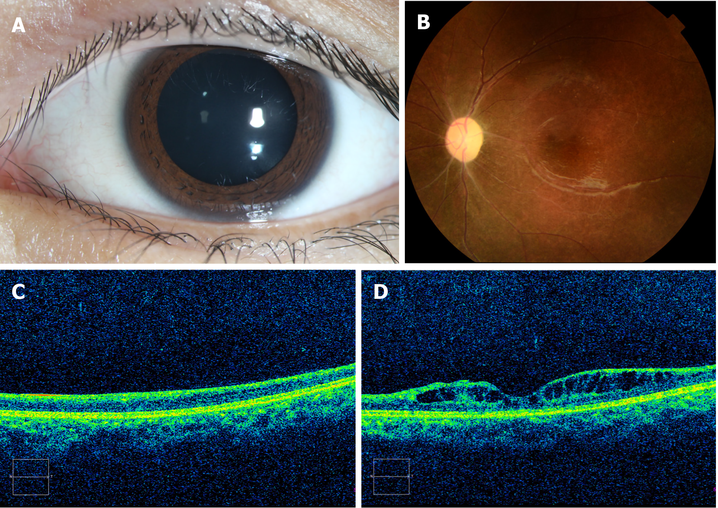
Figure 2 The girl’s ocular examination results.
A: The anterior segment was normal except for a relative afferent pupil defect, and the dilated pupil was drug-induced; B: Color fundus photographs showed a pale optic disc, retinal vascular occlusion and pigmentation in the peripheral retina; C and D: Optical coherence tomography images confirmed diffuse thinning of the whole retinal layer.
- Citation: Zheng XY, Cheng DJ, Lian LH, Zhang RT, Yu XY. Severe fundus lesions induced by ocular jellyfish stings: A case report. World J Clin Cases 2020; 8(19): 4544-4549
- URL: https://www.wjgnet.com/2307-8960/full/v8/i19/4544.htm
- DOI: https://dx.doi.org/10.12998/wjcc.v8.i19.4544


