Copyright
©The Author(s) 2020.
World J Clin Cases. Oct 6, 2020; 8(19): 4475-4480
Published online Oct 6, 2020. doi: 10.12998/wjcc.v8.i19.4475
Published online Oct 6, 2020. doi: 10.12998/wjcc.v8.i19.4475
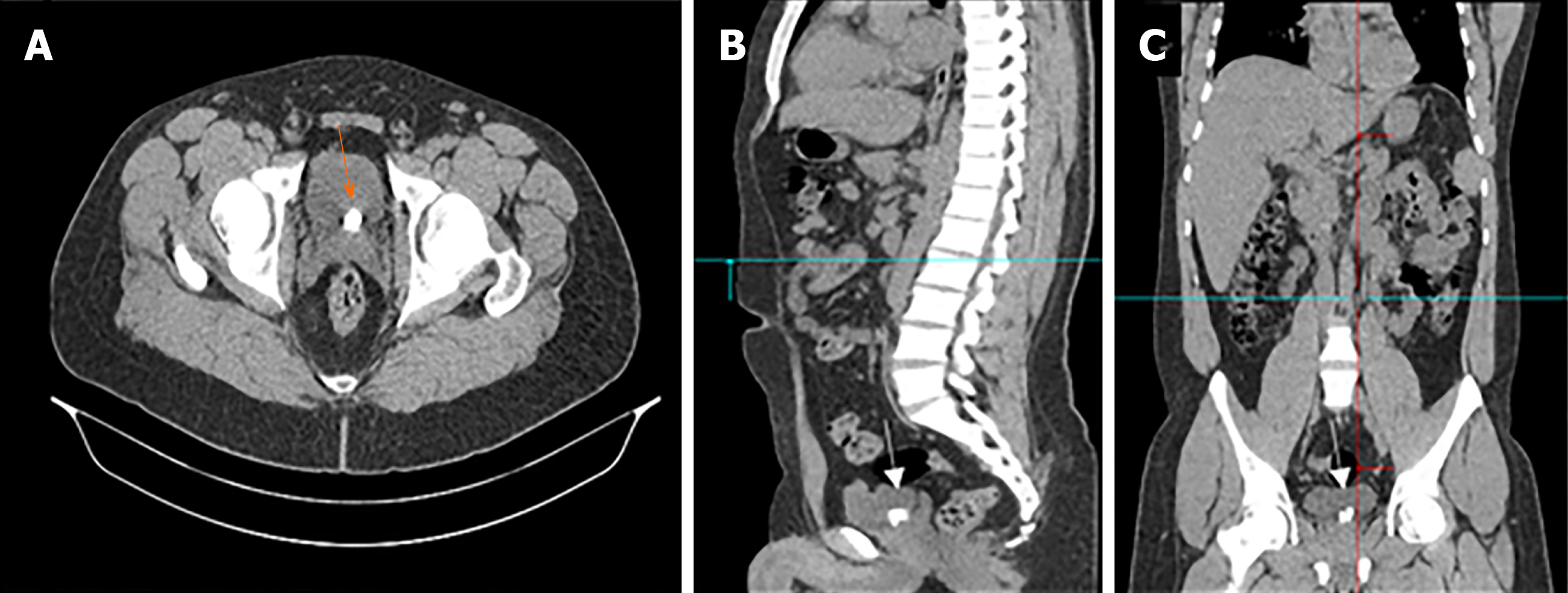
Figure 1 Computed tomography scan images showing two stones at the base of the bladder with a burden of 1.
5 cm. Computed tomography images do not reveal that the stones are present within a diverticulum (orange arrow). A: Axial; B: Sagittal; C: Coronal images.
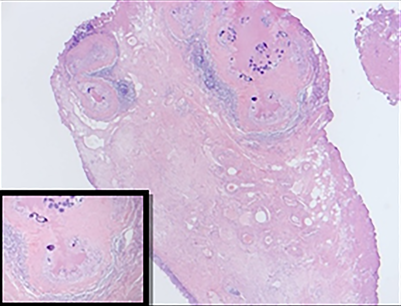
Figure 2 Hematoxylin and eosin staining images of resected diverticulum at 200× magnification.
Lamina propria show multiple granulomatous inflammation around calcified ova. Marked fibrosis and chronic inflammation were also observed surrounding the calcified ova.
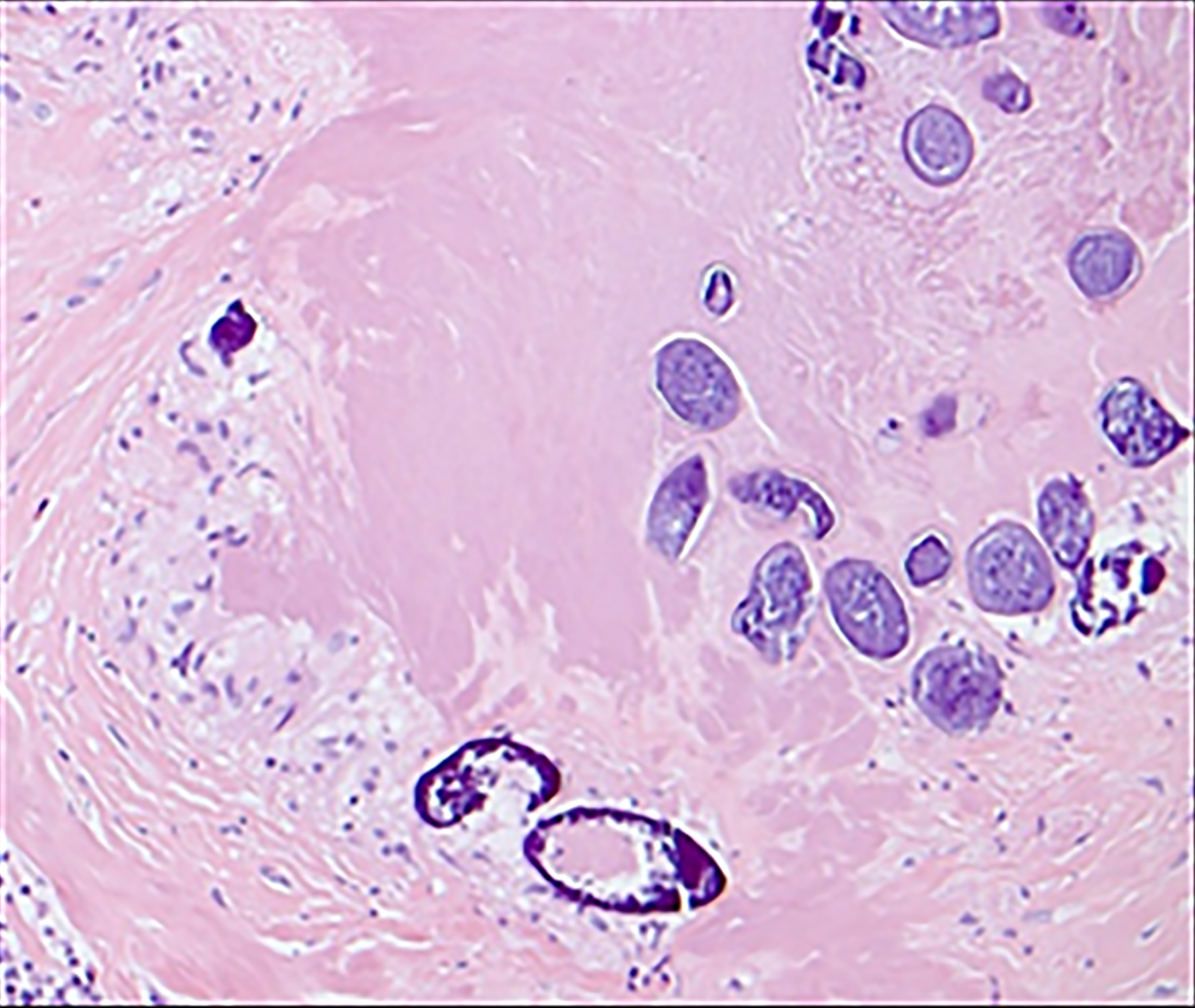
Figure 3 Hematoxylin and eosin staining images of resected diverticulum at 400× magnification.
The image shows that the majority of the Schistosoma eggs are calcified with some showing lateral spines.
- Citation: Alkhamees MA. Bladder stones in a closed diverticulum caused by Schistosoma mansoni: A case report. World J Clin Cases 2020; 8(19): 4475-4480
- URL: https://www.wjgnet.com/2307-8960/full/v8/i19/4475.htm
- DOI: https://dx.doi.org/10.12998/wjcc.v8.i19.4475


