Copyright
©The Author(s) 2020.
World J Clin Cases. Oct 6, 2020; 8(19): 4466-4474
Published online Oct 6, 2020. doi: 10.12998/wjcc.v8.i19.4466
Published online Oct 6, 2020. doi: 10.12998/wjcc.v8.i19.4466
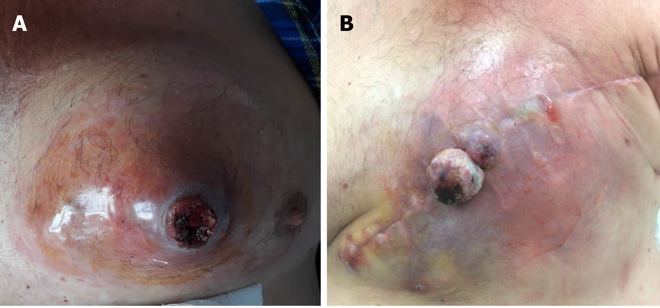
Figure 1 Macroscopic features.
A: Ulcerated rhabdomyosarcoma of the male breast; B: Tumor relapse, one month after mastectomy.
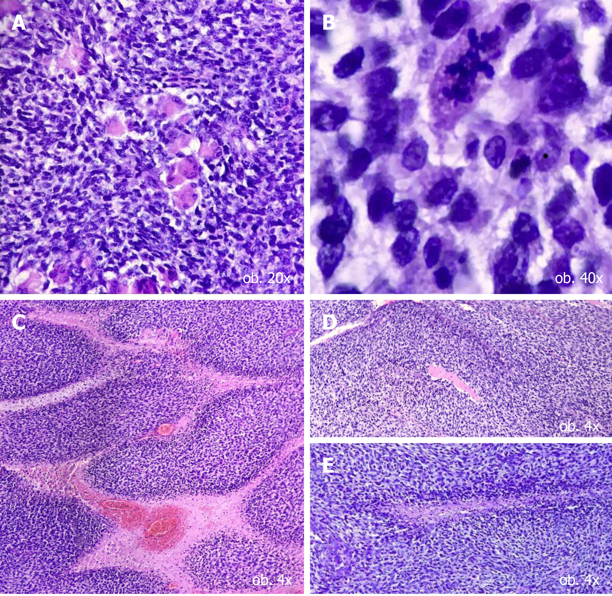
Figure 2 Histological features of a hybrid tumor of the breast.
A and B: Major population, some of the cells showing rhabdomyoblastic differentiation (A) and atypical mitoses (B); C-E: Small cell population, demonstrating lobular architecture (C), with formation of rosette-like structures (D and E).
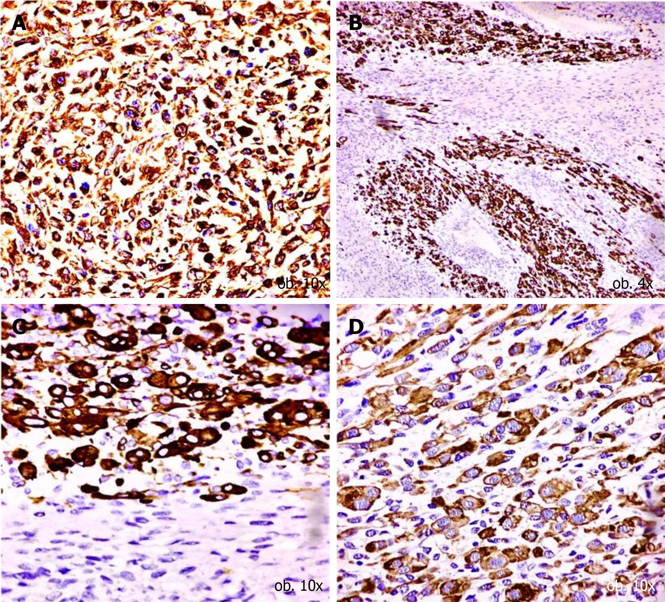
Figure 3 Immunohistochemical profile of major cell population.
A: Vimentin diffuse positivity confirms the mesenchymal origin; B-D: The rhabdomyosarcomatous differentiation is confirmed by the positivity for desmin (B), myogenin (C) and sarcomeric actin (D).
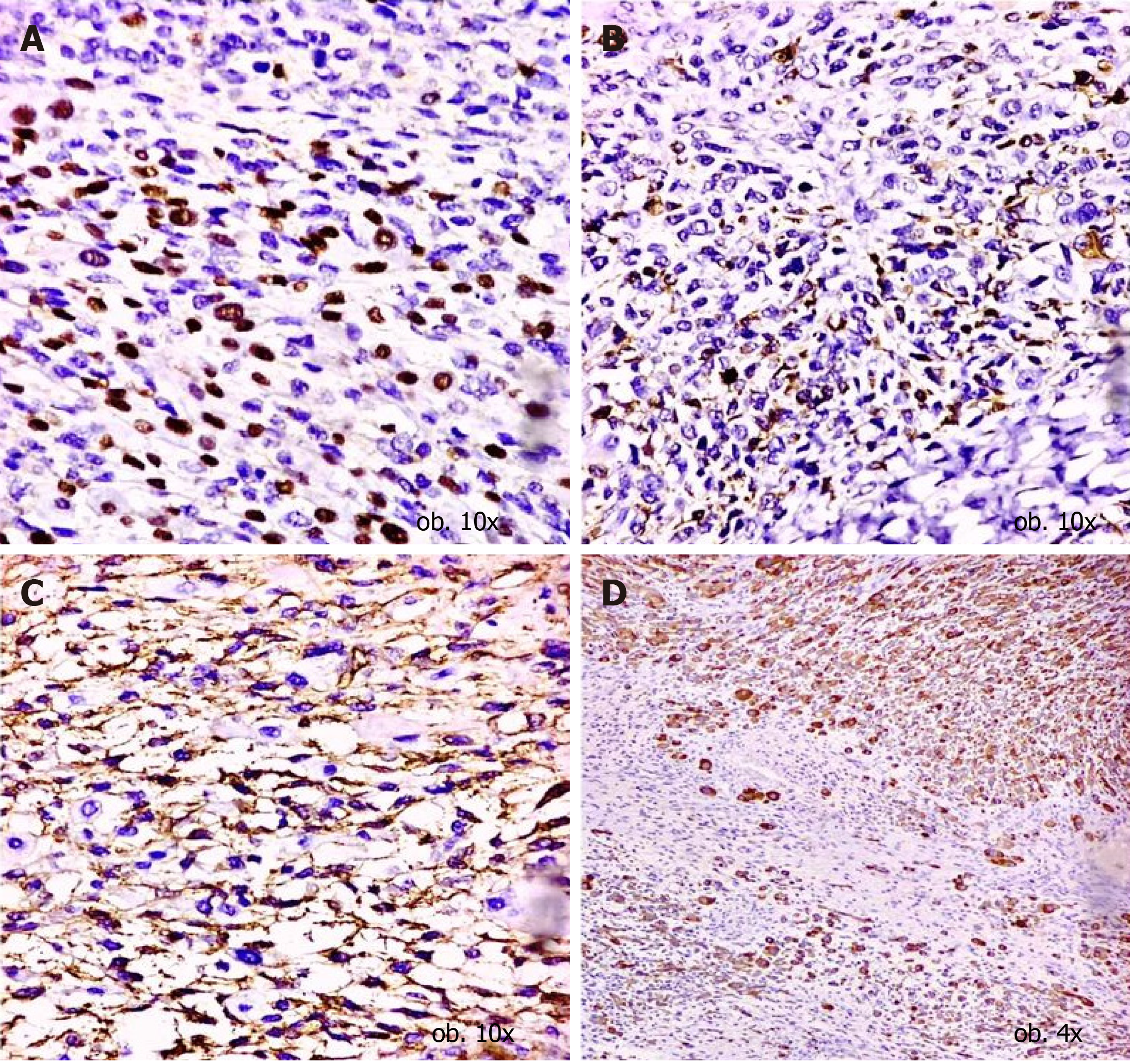
Figure 4 Immunohistochemical profile of small cell population.
A-C: The neuroectodermal origin is confirmed by the positivity for neurofilament (A), neuron specific enolase (B) and CD99 (C); D: CD56 positivity proves the interaction between myocytes and neural cells.
- Citation: Satală CB, Jung I, Bara TJ, Simu P, Simu I, Vlad M, Szodorai R, Gurzu S. Primary rhabdomyosarcoma: An extremely rare and aggressive variant of male breast cancer. World J Clin Cases 2020; 8(19): 4466-4474
- URL: https://www.wjgnet.com/2307-8960/full/v8/i19/4466.htm
- DOI: https://dx.doi.org/10.12998/wjcc.v8.i19.4466


