Copyright
©The Author(s) 2020.
World J Clin Cases. Oct 6, 2020; 8(19): 4400-4409
Published online Oct 6, 2020. doi: 10.12998/wjcc.v8.i19.4400
Published online Oct 6, 2020. doi: 10.12998/wjcc.v8.i19.4400
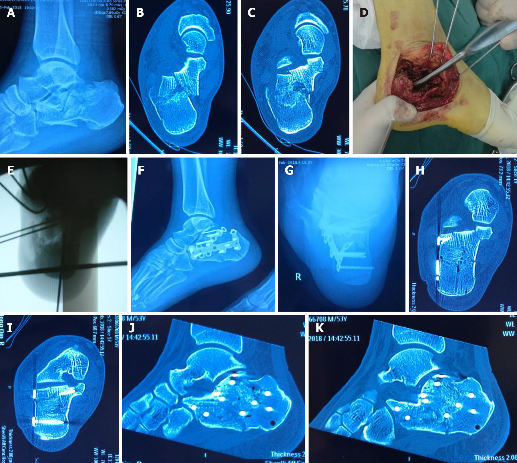
Figure 1 A 53-year-old man with fall injury and right calcaneal fracture, Sanders type II.
A-C: Preoperative images showing that the medial column was shortened, and the sustentaculum tali bone block and posterior nodule were tangled, which affected the reduction in the articular surface; D and E: The main fracture line was released during the operation, the medial column was reset, and the articular surface was reset after fixing with Kirschner wires; F-K: Postoperative images indicating that the medial column reduction was satisfactory, and the length, width, and force line of the calcaneus were effectively restored.
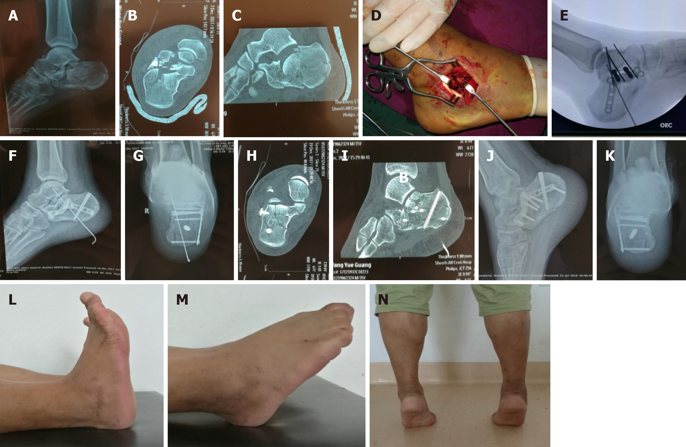
Figure 2 A 35-year-old man with fall injury and right calcaneal fracture, Sanders type III.
A-C: Preoperative images showing that the medial column was not shortened, and sustentaculum tali bone block and posterior nodule were not tangled, which would not affect the reduction of the articular surface; D and E: The articular surface bone block was reset from the inside to the outside during operation through the tarsal sinus incision; F-I: Postoperative images showing articular surface smoothing; J-N: Fractures healed at 4 mo after operation and function recovery was satisfactory.
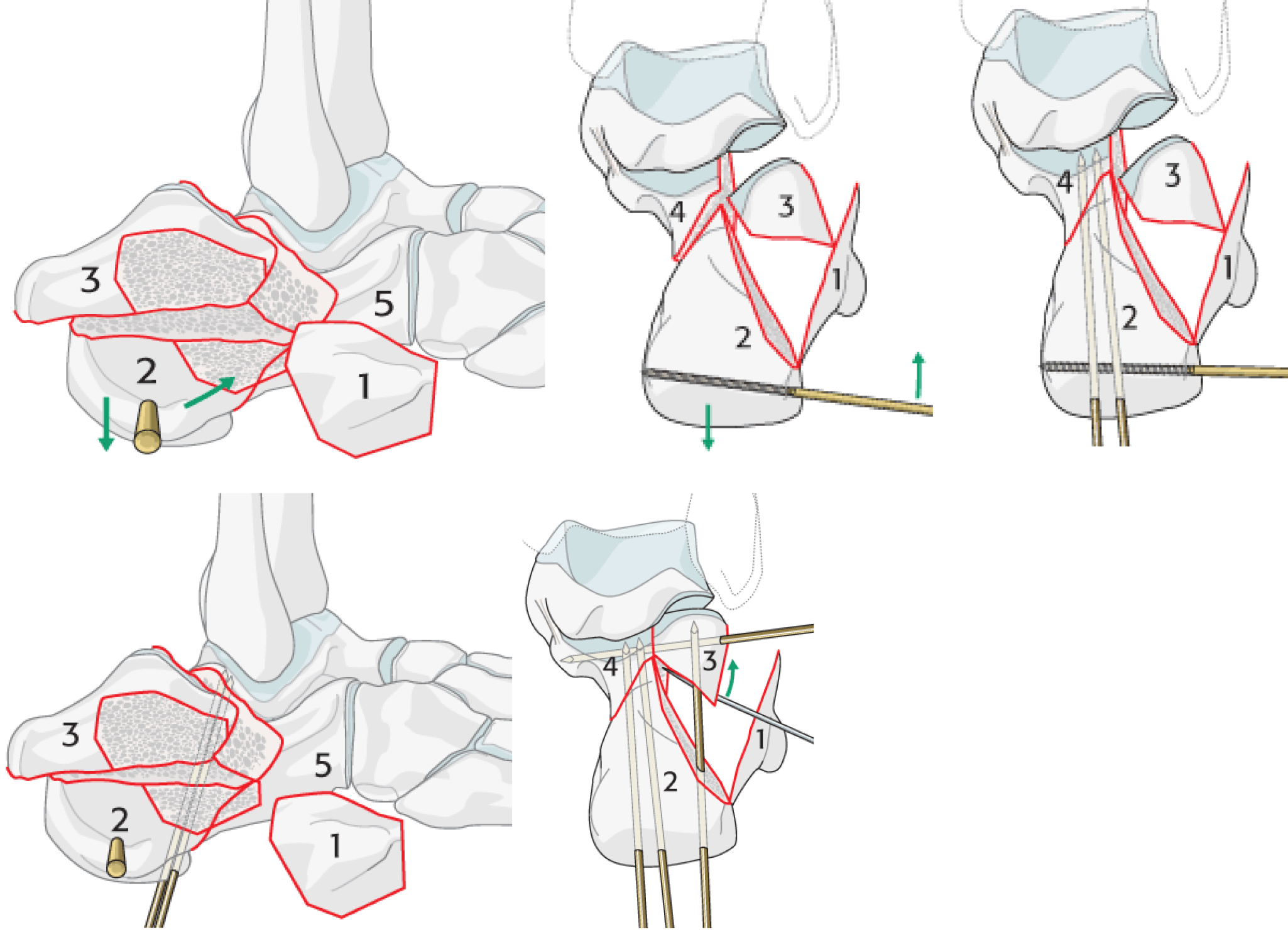
Figure 3 Reduction procedure for calcaneal fractures recommended by AO Course.
- Citation: Zheng G, Xia F, Yang S, Cui J. Application of medial column classification in treatment of intra-articular calcaneal fractures. World J Clin Cases 2020; 8(19): 4400-4409
- URL: https://www.wjgnet.com/2307-8960/full/v8/i19/4400.htm
- DOI: https://dx.doi.org/10.12998/wjcc.v8.i19.4400


