Copyright
©The Author(s) 2020.
World J Clin Cases. May 26, 2020; 8(10): 1950-1957
Published online May 26, 2020. doi: 10.12998/wjcc.v8.i10.1950
Published online May 26, 2020. doi: 10.12998/wjcc.v8.i10.1950
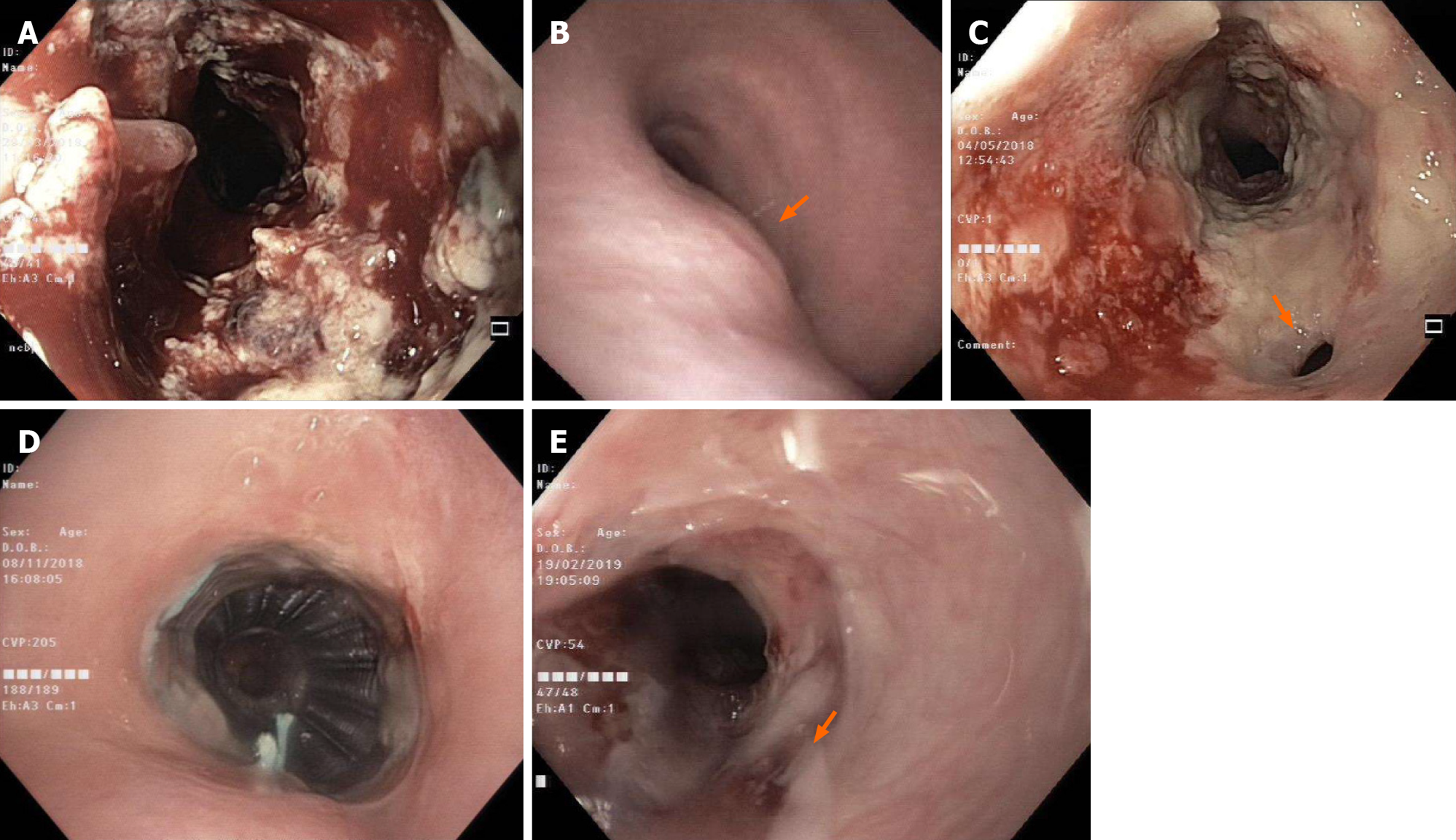
Figure 1 Endoscopic and imaging examinations.
A and B: Esophageal tumor on esophagogastroduodenocopy (A) and airway evaluation on bronchoscopy (B) where an extrinsic compression of trachea (arrow) was observed at initial diagnosis; C: A tracheoesophageal fistula (arrow) seen on esophagogastroduodenocopy after first week of chemoradiotherapy; D: Tracheal stent in place on bronchoscopy at 6-mo post-chemoradiotherapy; E: Complete tumor response with closure of fistula (arrow) evident on esophagogastroduodenocopy at 6-mo post-chemoradiotherapy.
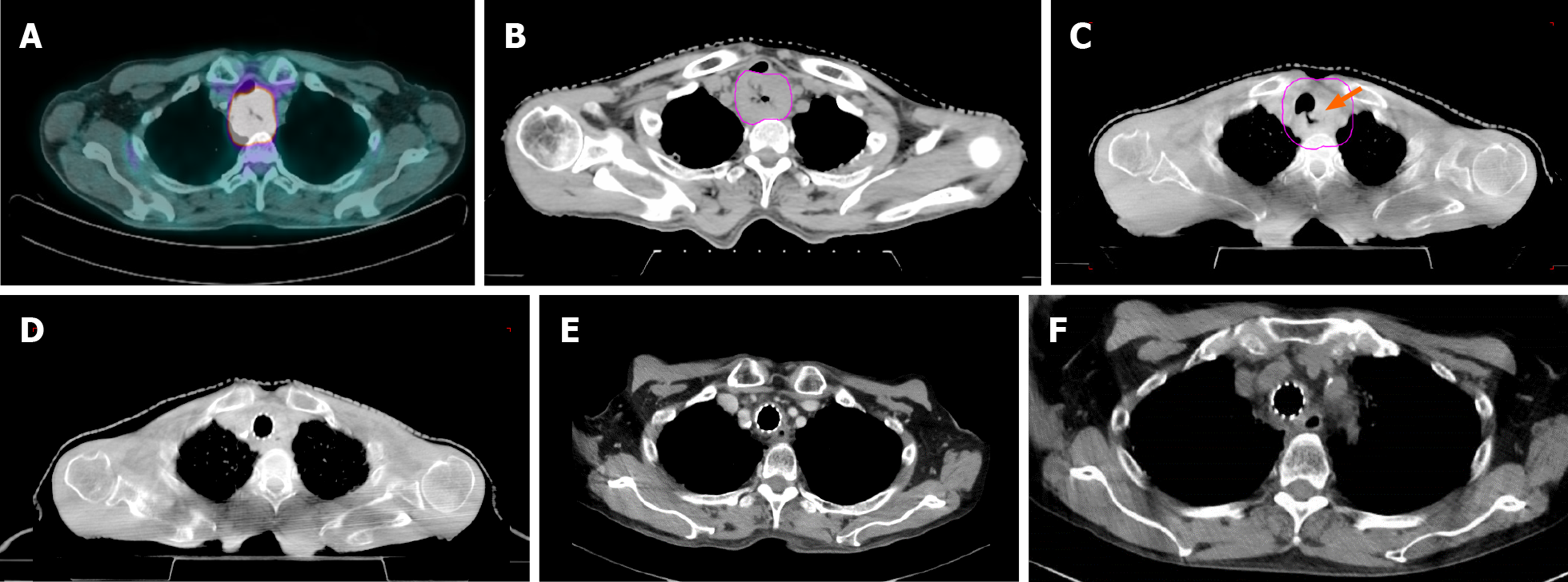
Figure 2 Radiographic findings of the tumor.
A: Positron emission tomography-computed tomography (CT) at initial diagnosis; B-D: Cone-beam CT on first fraction (B), fifth fraction where tumor response was evident with the development of tracheoesophageal fistula (arrow) (C), and final fraction of radiotherapy when fistula was closed (D); E: Contrasted CT imaging at 6-mo post-chemoradiotherapy showing complete response; F: Contrasted CT imaging at 15-mo post-chemoradiotherapy showing no recurrence.
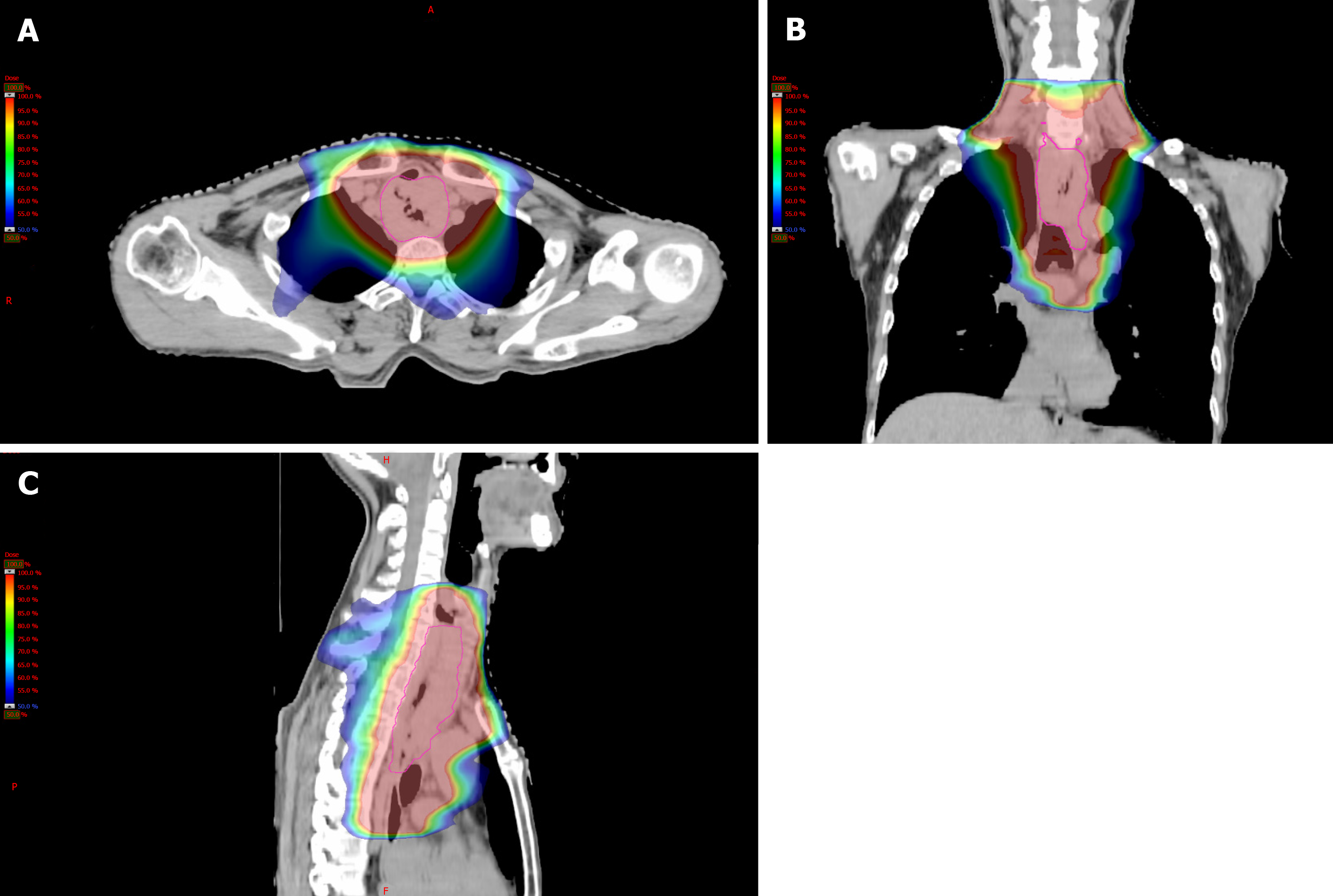
Figure 3 Radiation plan using intensity-modulated radiation therapy technique.
A: Axial view; B: Coronal view; and C: Sagittal view.
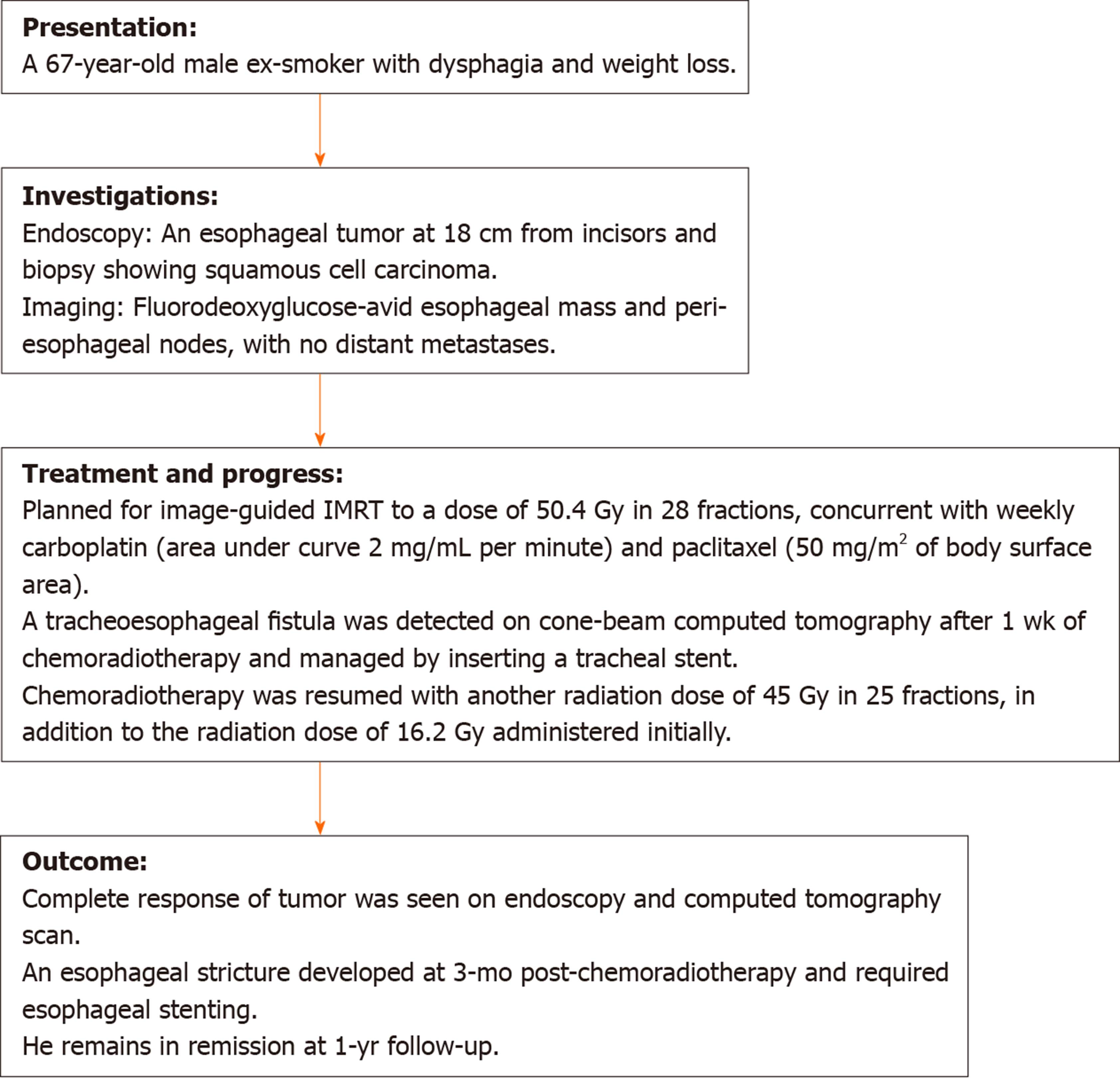
Figure 4 Timeline of events.
IMRT: Intensity-modulated radiation therapy.
- Citation: Lee CC, Yeo CM, Ng WK, Verma A, Tey JC. T4 cervical esophageal cancer cured with modern chemoradiotherapy: A case report. World J Clin Cases 2020; 8(10): 1950-1957
- URL: https://www.wjgnet.com/2307-8960/full/v8/i10/1950.htm
- DOI: https://dx.doi.org/10.12998/wjcc.v8.i10.1950


