Copyright
©The Author(s) 2019.
World J Clin Cases. May 6, 2019; 7(9): 1080-1086
Published online May 6, 2019. doi: 10.12998/wjcc.v7.i9.1080
Published online May 6, 2019. doi: 10.12998/wjcc.v7.i9.1080
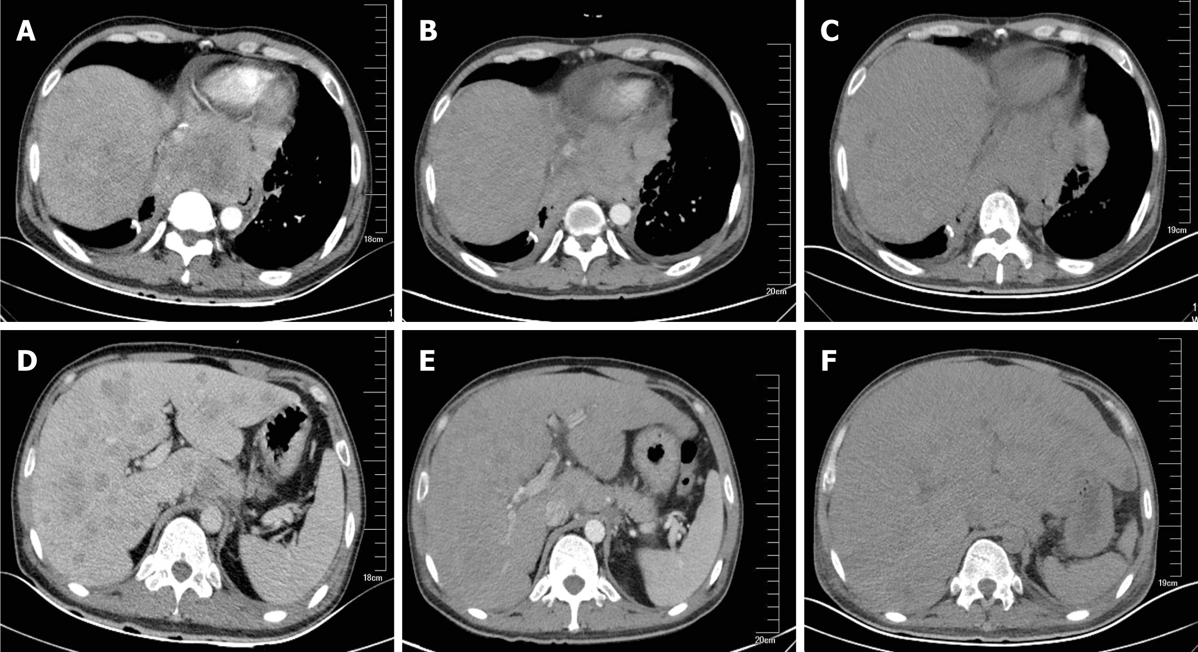
Figure 1 Computed tomography indicated mediastinal and hepatic metastatic changes.
A: Chest computed tomography (CT) showed a 8.4 × 9.8 cm mediastinal metastatic lump; B: Chest CT on day 35 showed a slight shrink in mediastinal metastasis; C: Chest CT on day 55 showed unchanged mediastinal lump compared to B; D: Abdominal CT showed multiple metastases; E: Liver metastasis slightly shrank after 35 d of crizotinib therapy; F: Liver volume increased by 20% and acute intrahepatic bile were revealed on day 55.
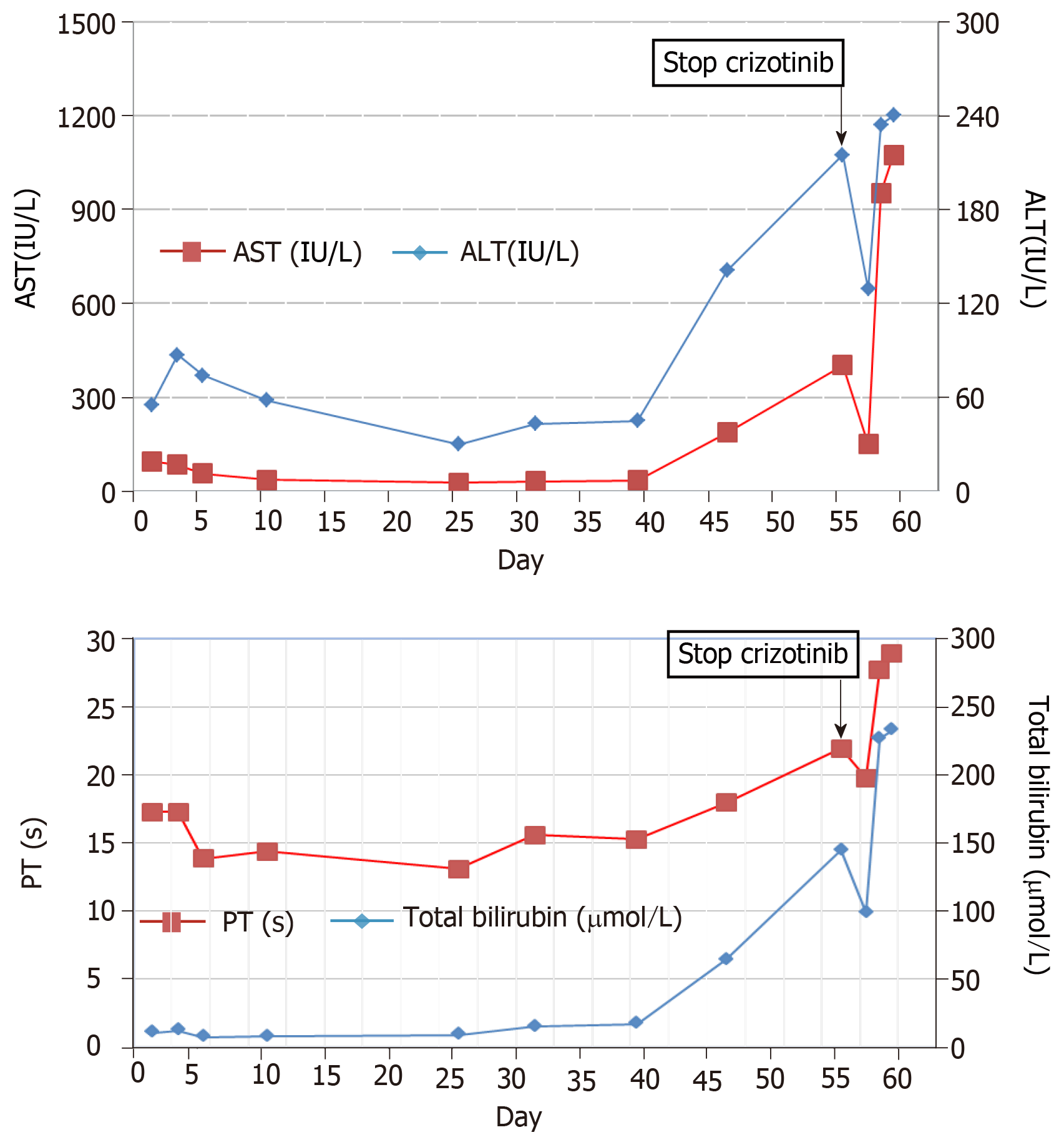
Figure 2 Detailed changes of liver enzymes during crizotinib therapy (day 1–55) and after crizotinib discontinuation (day 56–59).
A: Upper graph: aspartate aminotransferase and alanine aminotransferase; B: Lower graph: total bilirubin and prothrombin time. ALT: Alanine aminotransferase; AST: Aspartate aminotransferase; PT: Prothrombin time.
- Citation: Zhang Y, Xu YY, Chen Y, Li JN, Wang Y. Crizotinib-induced acute fatal liver failure in an Asian ALK-positive lung adenocarcinoma patient with liver metastasis: A case report. World J Clin Cases 2019; 7(9): 1080-1086
- URL: https://www.wjgnet.com/2307-8960/full/v7/i9/1080.htm
- DOI: https://dx.doi.org/10.12998/wjcc.v7.i9.1080


