Copyright
©The Author(s) 2019.
World J Clin Cases. Apr 26, 2019; 7(8): 951-960
Published online Apr 26, 2019. doi: 10.12998/wjcc.v7.i8.951
Published online Apr 26, 2019. doi: 10.12998/wjcc.v7.i8.951
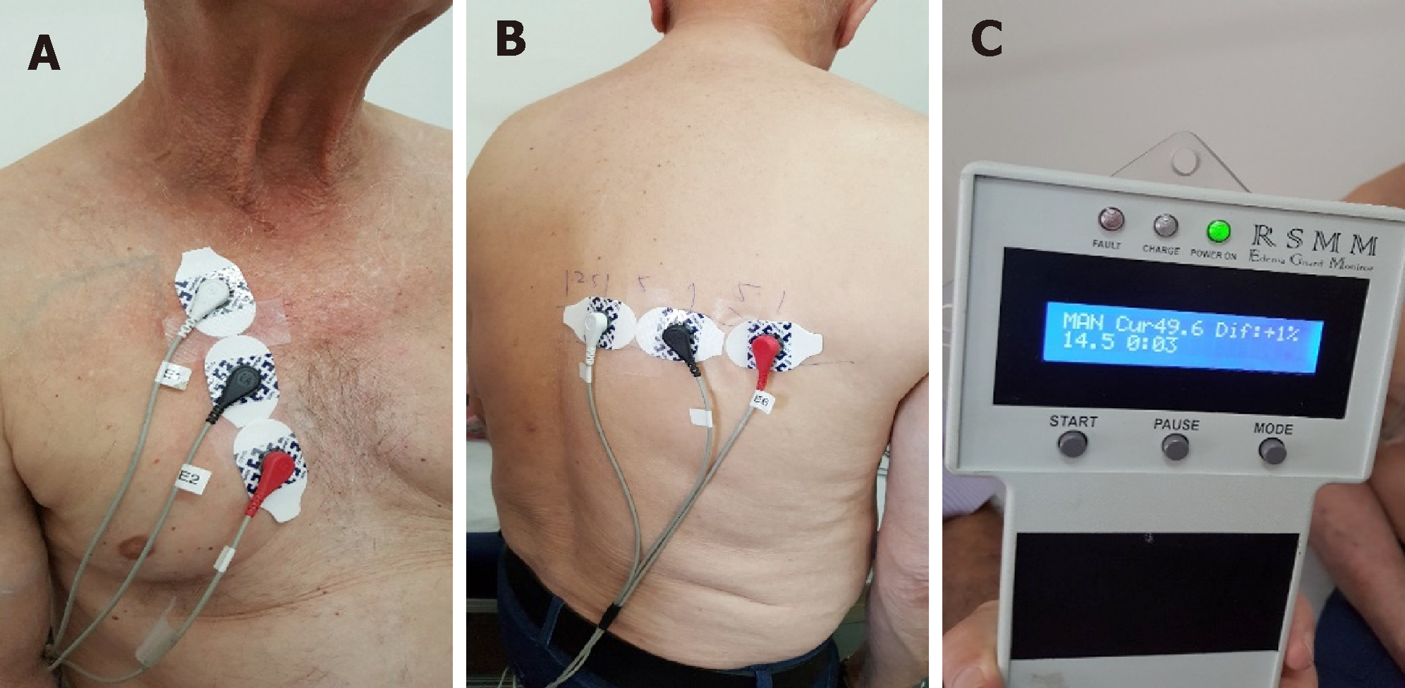
Figure 1 The technique of lung impedance measurements.
A: Three electrodes were placed vertically on the front right side of the chest, 4.5 cm from the midline of the sternum with the upper electrode attached precisely under the clavicle; B: Another set of 3 electrodes was placed on the back along the horizontal line crossing the low edge of the right scapula, with the most leftward electrode placed at the crossing point of the horizontal line with the spine; C: The LI measurement result was displayed as a number on the screen.
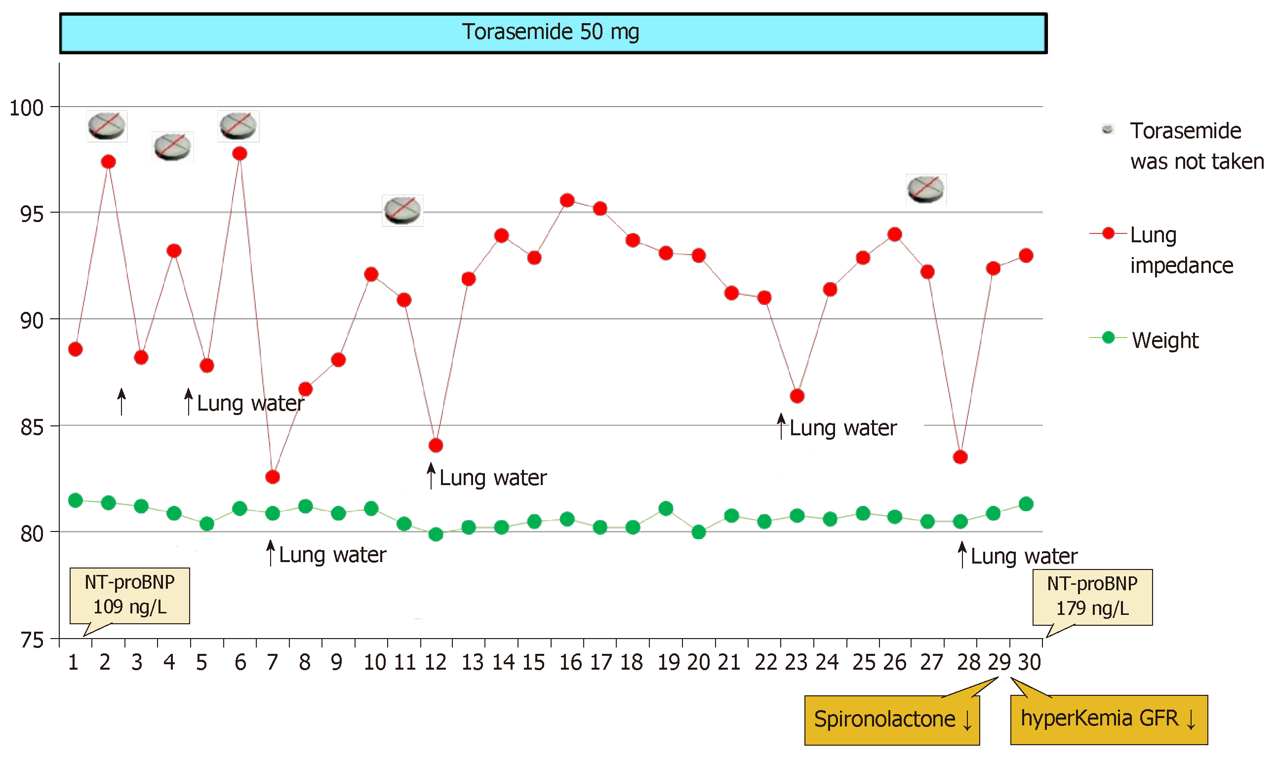
Figure 2 The dynamics of lung impedance, weight and diuretic dose for Patient 1.
The N-terminal pro B-type natriuretic peptide level was not high at the day of discharge, but the patient had clear signs and symptoms of congestive heart failure (HF) on admission (shortness of breath, bilateral rales in the lungs, leg edema, poor left ventricular function on echocardiography). HF with reduced ejection fraction developed after an acute myocardial infarction more than 4 years ago; then, his BNP was 2611 ng/L, but 2 years after, it had reduced to the level of 200 ng/L. NT-proBNP: N-terminal pro B-type natriuretic peptide; GFR: Glomerular filtration rate.
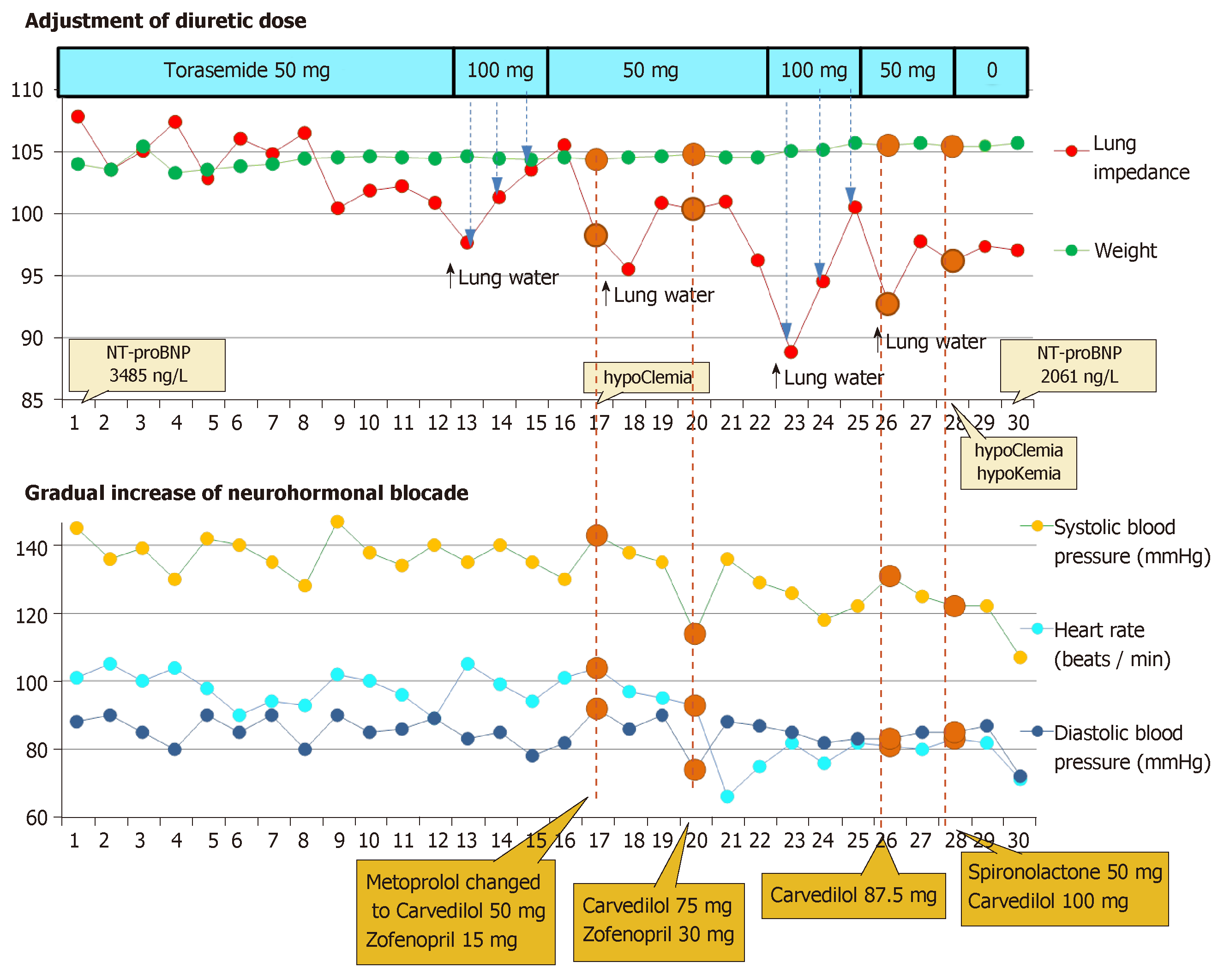
Figure 3 The dynamics of lung congestion, weight, hemodynamics and medication dosage for Patient 2.
NT-proBNP: N-terminal pro B-type natriuretic peptide.
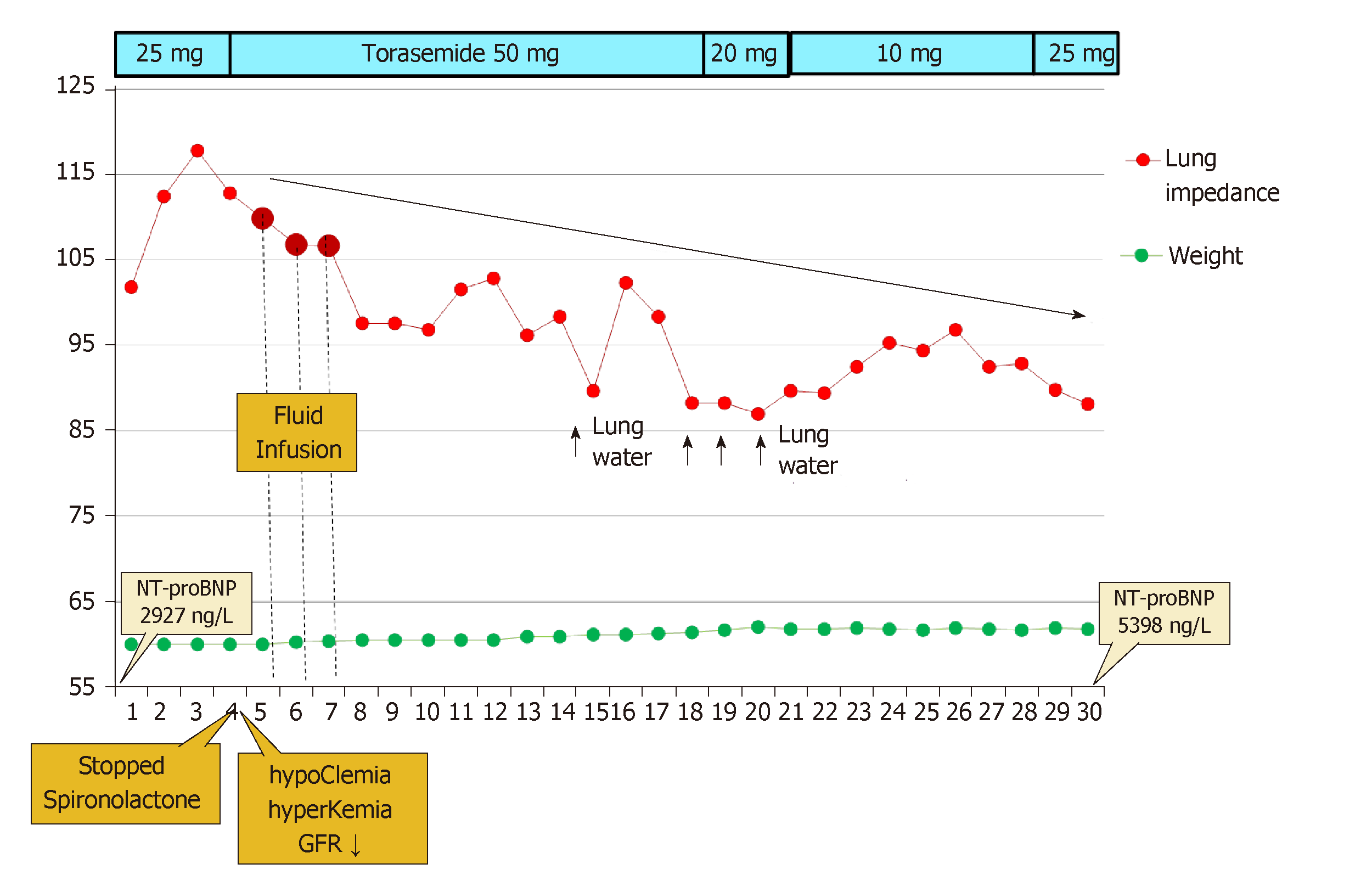
Figure 4 The dynamics of lung congestion, weight and diuretic dose for Patient 3 (in this figure, the legend is different than the others (LI).
NT-proBNP: N-terminal pro B-type natriuretic peptide; GFR: Glomerular filtration rate.
- Citation: Lycholip E, Palevičiūtė E, Aamodt IT, Hellesø R, Lie I, Strömberg A, Jaarsma T, Čelutkienė J. Non-invasive home lung impedance monitoring in early post-acute heart failure discharge: Three case reports. World J Clin Cases 2019; 7(8): 951-960
- URL: https://www.wjgnet.com/2307-8960/full/v7/i8/951.htm
- DOI: https://dx.doi.org/10.12998/wjcc.v7.i8.951


