Copyright
©The Author(s) 2019.
World J Clin Cases. Apr 6, 2019; 7(7): 903-907
Published online Apr 6, 2019. doi: 10.12998/wjcc.v7.i7.903
Published online Apr 6, 2019. doi: 10.12998/wjcc.v7.i7.903
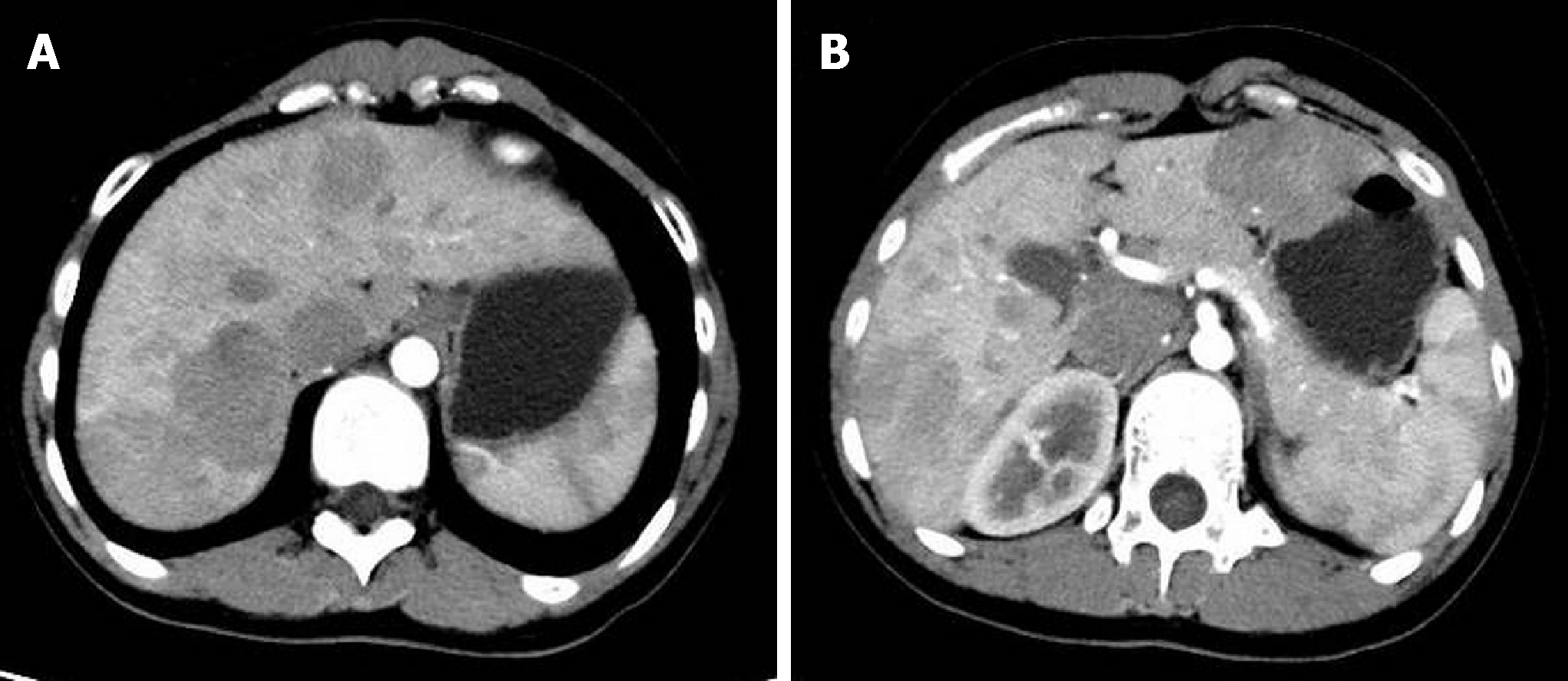
Figure 1 Computed tomography arterial phase.
Several hypovascular masses are seen in the right hemiliver (A); and the left hemiliver (B).
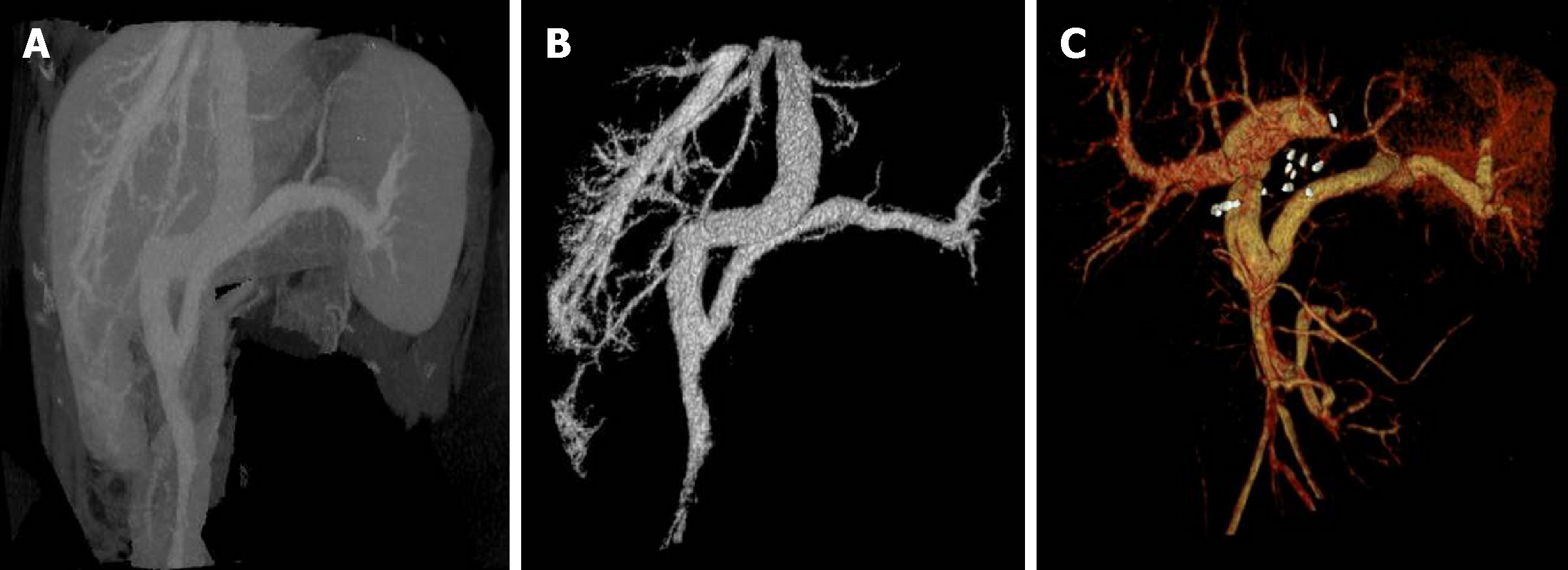
Figure 2 Computed tomography 3D reconstruction and portal vein image reconstruction.
A, B: Pre-operation computed tomography 3D reconstruction and portal vein image reconstruction show that the superior mesenteric vein and splenic vein converge to form the portal vein, and the portal vein directly enters the inferior vena cava; C: post-operation computed tomography portal vein image reconstruction shows the liver transplantation corrects the patient's anatomical abnormalities.
- Citation: Xiang W, Wang H, Si ZZ, Chen GS, Wang GW, Li T. Type I congenital extrahepatic portosystemic shunt treated by orthotopic liver transplantation: A case report. World J Clin Cases 2019; 7(7): 903-907
- URL: https://www.wjgnet.com/2307-8960/full/v7/i7/903.htm
- DOI: https://dx.doi.org/10.12998/wjcc.v7.i7.903


