Copyright
©The Author(s) 2019.
World J Clin Cases. Apr 6, 2019; 7(7): 903-907
Published online Apr 6, 2019. doi: 10.12998/wjcc.v7.i7.903
Published online Apr 6, 2019. doi: 10.12998/wjcc.v7.i7.903
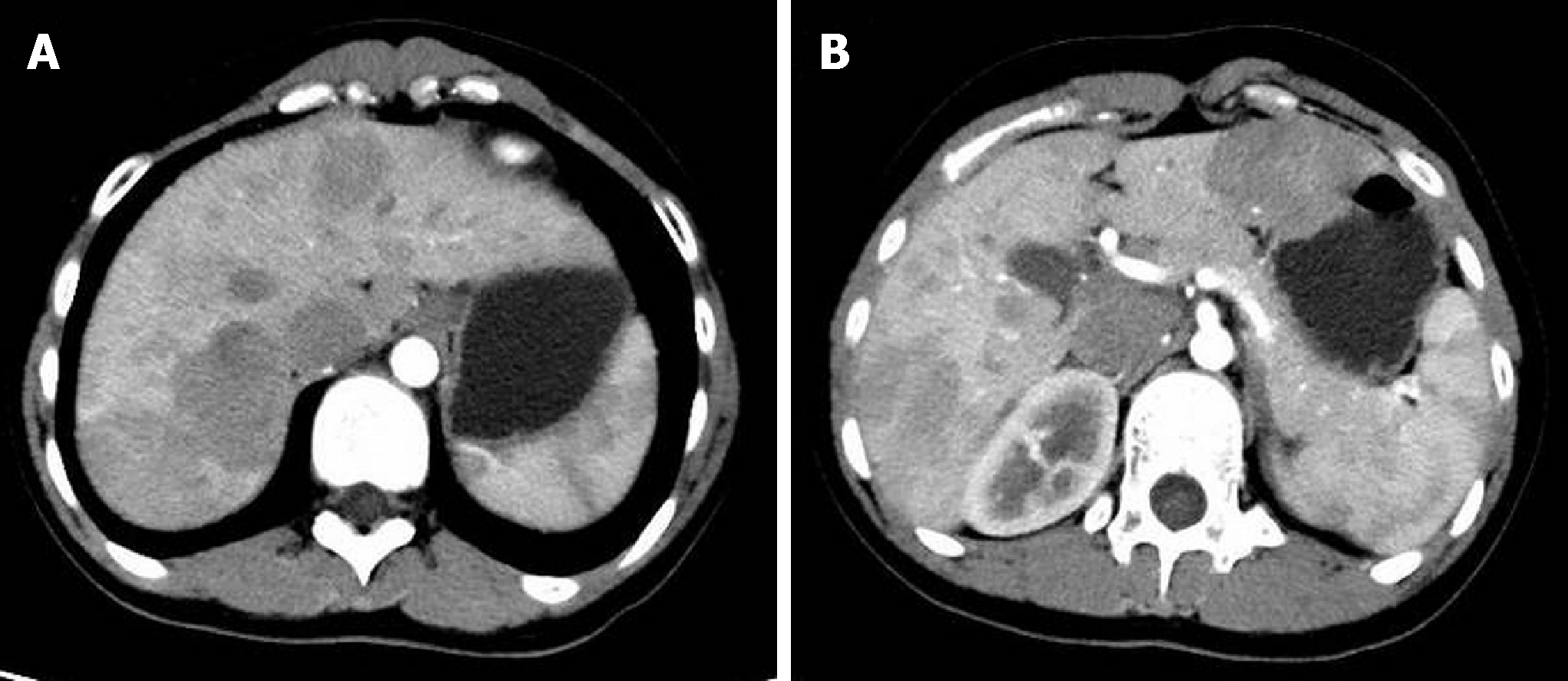
Figure 1 Computed tomography arterial phase.
Several hypovascular masses are seen in the right hemiliver (A); and the left hemiliver (B).
- Citation: Xiang W, Wang H, Si ZZ, Chen GS, Wang GW, Li T. Type I congenital extrahepatic portosystemic shunt treated by orthotopic liver transplantation: A case report. World J Clin Cases 2019; 7(7): 903-907
- URL: https://www.wjgnet.com/2307-8960/full/v7/i7/903.htm
- DOI: https://dx.doi.org/10.12998/wjcc.v7.i7.903


