Copyright
©The Author(s) 2019.
World J Clin Cases. Mar 26, 2019; 7(6): 753-758
Published online Mar 26, 2019. doi: 10.12998/wjcc.v7.i6.753
Published online Mar 26, 2019. doi: 10.12998/wjcc.v7.i6.753
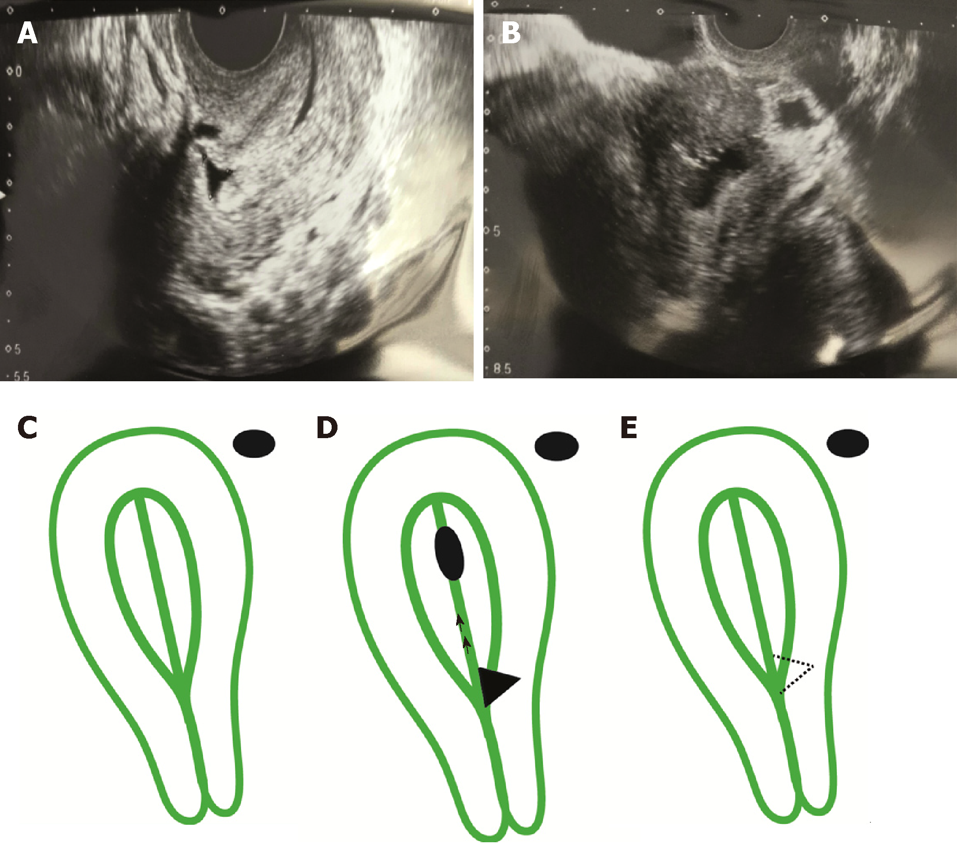
Figure 1 Transvaginal ultrasonographic images.
A 37-year-old female suffering from persistent hydrometra during in vitro fertilization underwent a transvaginal ultrasonographic examination. A: Panel A indicates the presence of a second-grade isthmocele; size 6.6 mm (base) × 6.1 mm (height); B: Panel B demonstrates the hydrometra and isthmocele. All images were taken with a Toshiba Xario 100 ultrasound with an endovaginal probe, frequency 7 megahertz (MHz); C: Diagrammatic representations of the uterus showing a normal uterus in Panel C; D: Hydrometra and isthmocele in Panel D; E: and result after resection in Panel E.
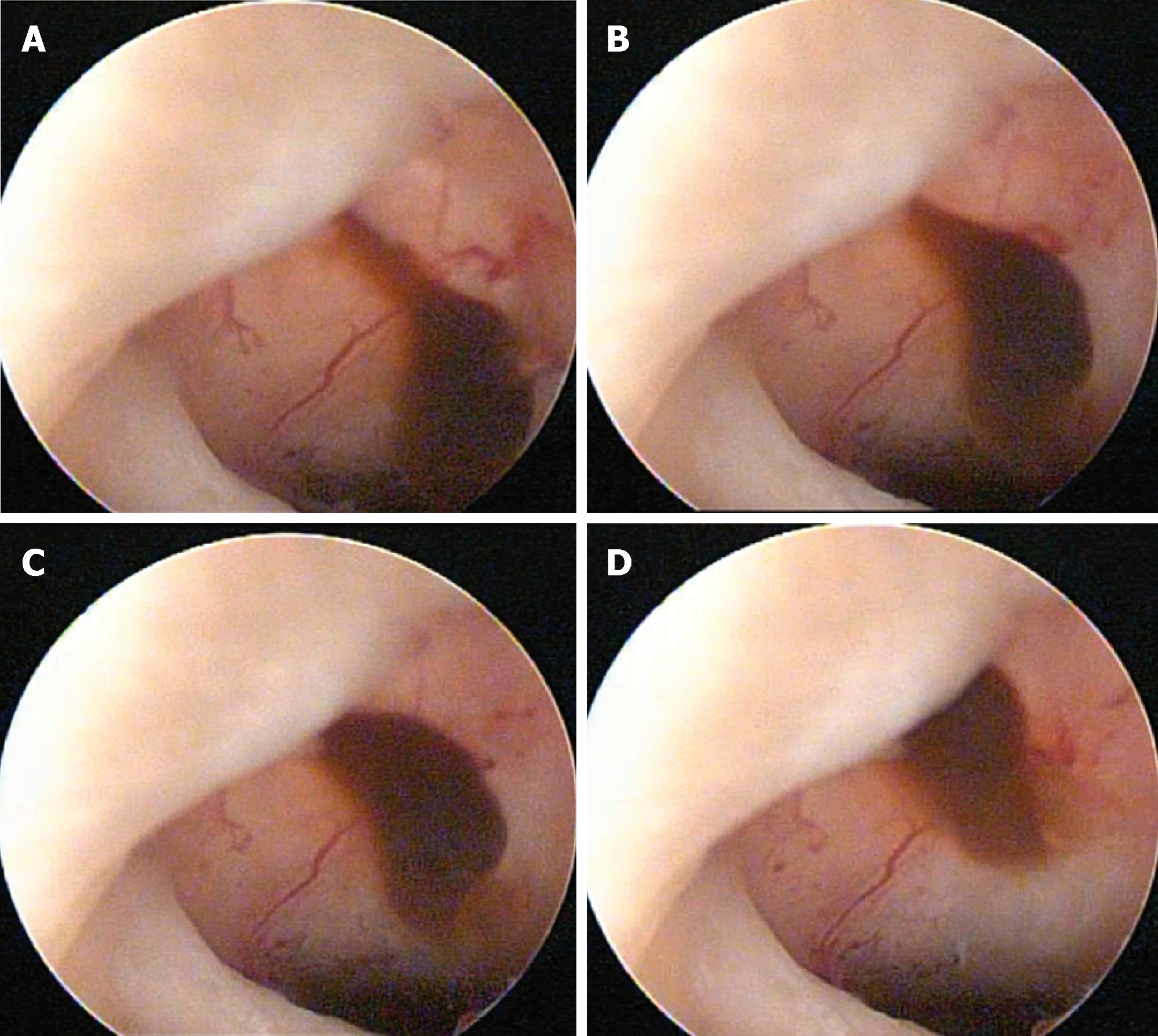
Figure 2 Isthmocele at the time of surgery.
Images were selected from the video recorded during surgery. The procedure was performed by finding the myocell sac containing neovascularization and mucosanguineous content, leveling the isthmocele area with a monopolar resectoscope loop, and subsequent application of monopolar ablation energy in the bed of the isthmocele using a roll ball electrode. The procedure was performed guided at times by transabdominal ultrasound (procedure time: 25 min; 2× magnification).
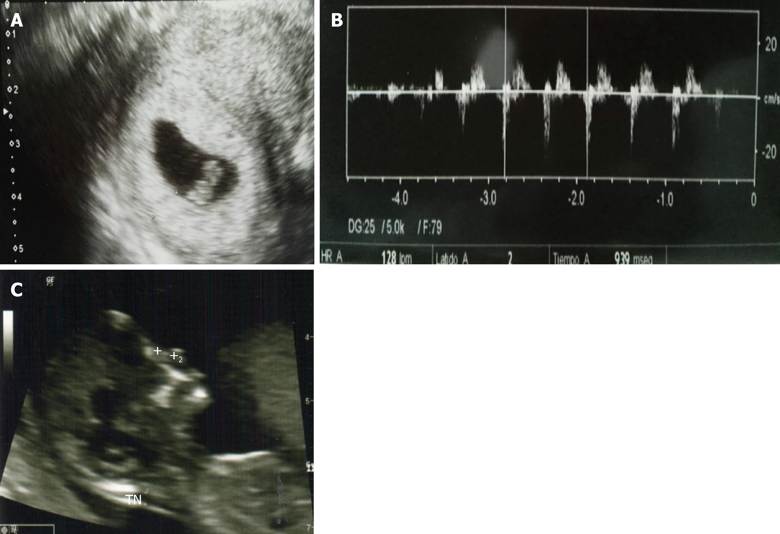
Figure 3 Pregnancy ultrasounds.
After isthmocele correction, 2 embryos were implanted. A: Panel A indicates that at Week 6 the presence of one fetal sac by transvaginal ultrasonogram; B: Transabdominal ecogram is showing the embryo’s heartbeat; C: Panel C is showing the live fetus, normal nasal bond (2.5 cm), normal nuchal fold thickening (1.8), and no signs of chromosomopathies, using transabdominal ultrasonogram. The length of the cervix was 3.3 cm.
- Citation: López Rivero LP, Jaimes M, Camargo F, López-Bayghen E. Successful treatment with hysteroscopy for infertility due to isthmocele and hydrometra secondary to cesarean section: A case report. World J Clin Cases 2019; 7(6): 753-758
- URL: https://www.wjgnet.com/2307-8960/full/v7/i6/753.htm
- DOI: https://dx.doi.org/10.12998/wjcc.v7.i6.753


