
















©The Author(s) 2019.
World J Clin Cases. Mar 26, 2019; 7(6): 753-758
Published online Mar 26, 2019. doi: 10.12998/wjcc.v7.i6.753
Published online Mar 26, 2019. doi: 10.12998/wjcc.v7.i6.753
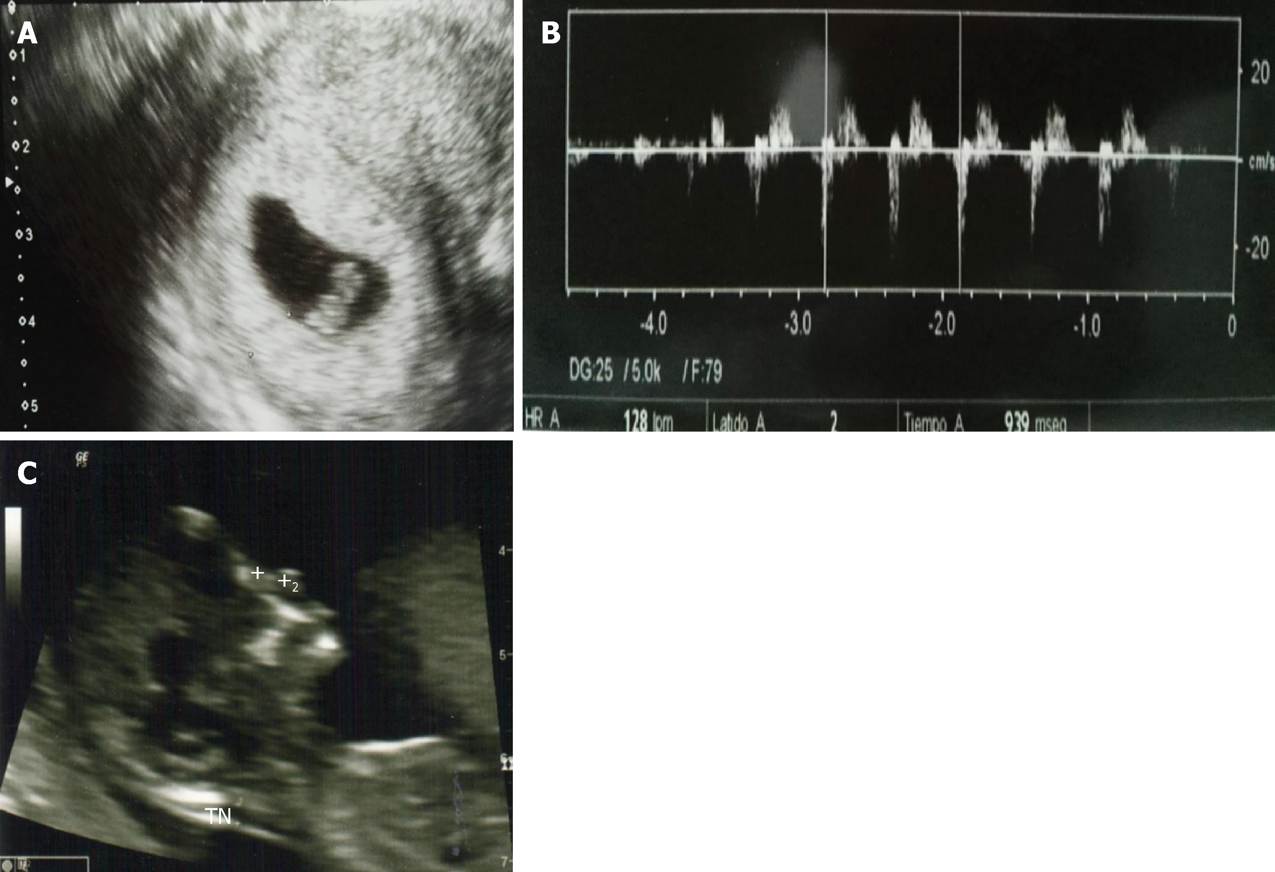
Figure 3 Pregnancy ultrasounds.
After isthmocele correction, 2 embryos were implanted. A: Panel A indicates that at Week 6 the presence of one fetal sac by transvaginal ultrasonogram; B: Transabdominal ecogram is showing the embryo’s heartbeat; C: Panel C is showing the live fetus, normal nasal bond (2.5 cm), normal nuchal fold thickening (1.8), and no signs of chromosomopathies, using transabdominal ultrasonogram. The length of the cervix was 3.3 cm.
- Citation: López Rivero LP, Jaimes M, Camargo F, López-Bayghen E. Successful treatment with hysteroscopy for infertility due to isthmocele and hydrometra secondary to cesarean section: A case report. World J Clin Cases 2019; 7(6): 753-758
- URL: https://www.wjgnet.com/2307-8960/full/v7/i6/753.htm
- DOI: https://dx.doi.org/10.12998/wjcc.v7.i6.753