Copyright
©The Author(s) 2019.
World J Clin Cases. Dec 6, 2019; 7(23): 4130-4136
Published online Dec 6, 2019. doi: 10.12998/wjcc.v7.i23.4130
Published online Dec 6, 2019. doi: 10.12998/wjcc.v7.i23.4130
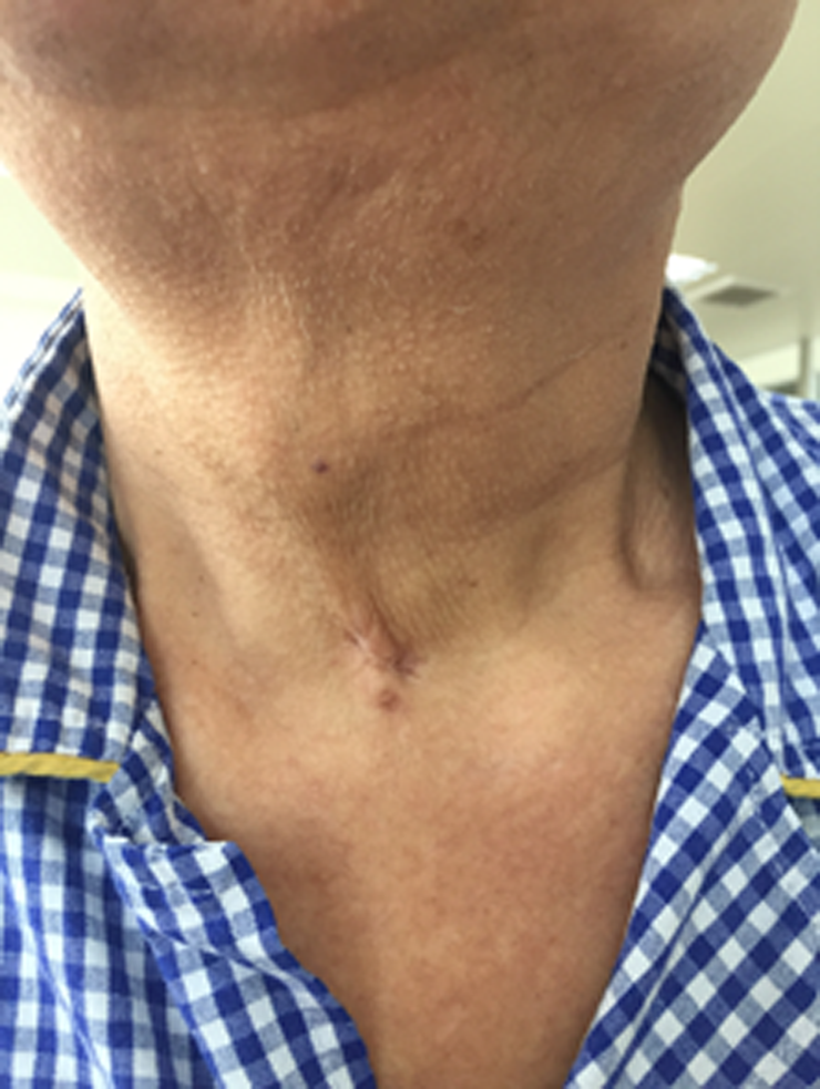
Figure 1 Wound healing with a scar formation on the anterior neck.
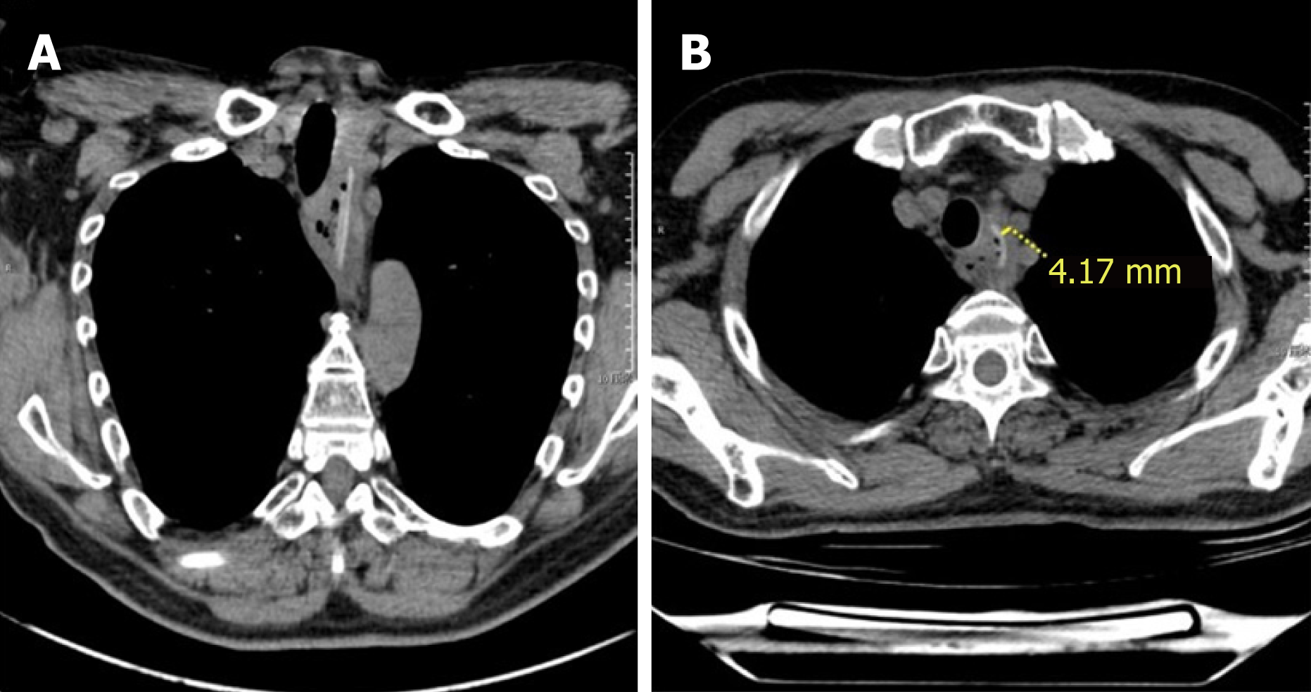
Figure 2 Locations of the embedded foreign bodies.
A: Chest coronal computed tomography image showing two paratactic foreign bodies penetrating from the superior mediastinal to the esophagus diagonally; B: Chest axial computed tomography image showing the upper ends of the foreign bodies outside the esophagus, adjacent to the left subclavian artery (4.1 mm).
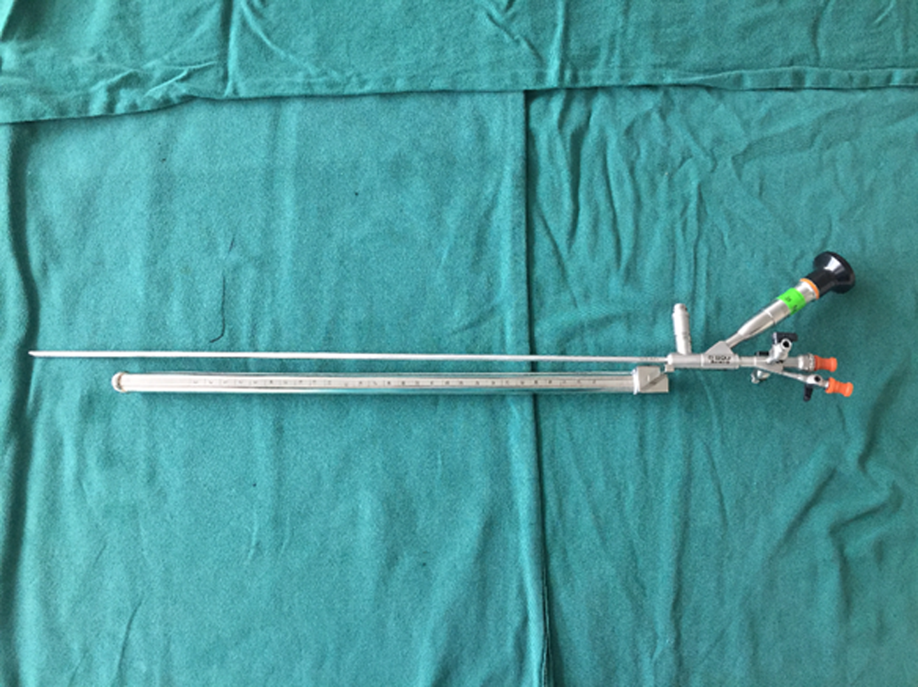
Figure 3 Rigid esophagoscope combined with an angle endoscope (ureteroscope).
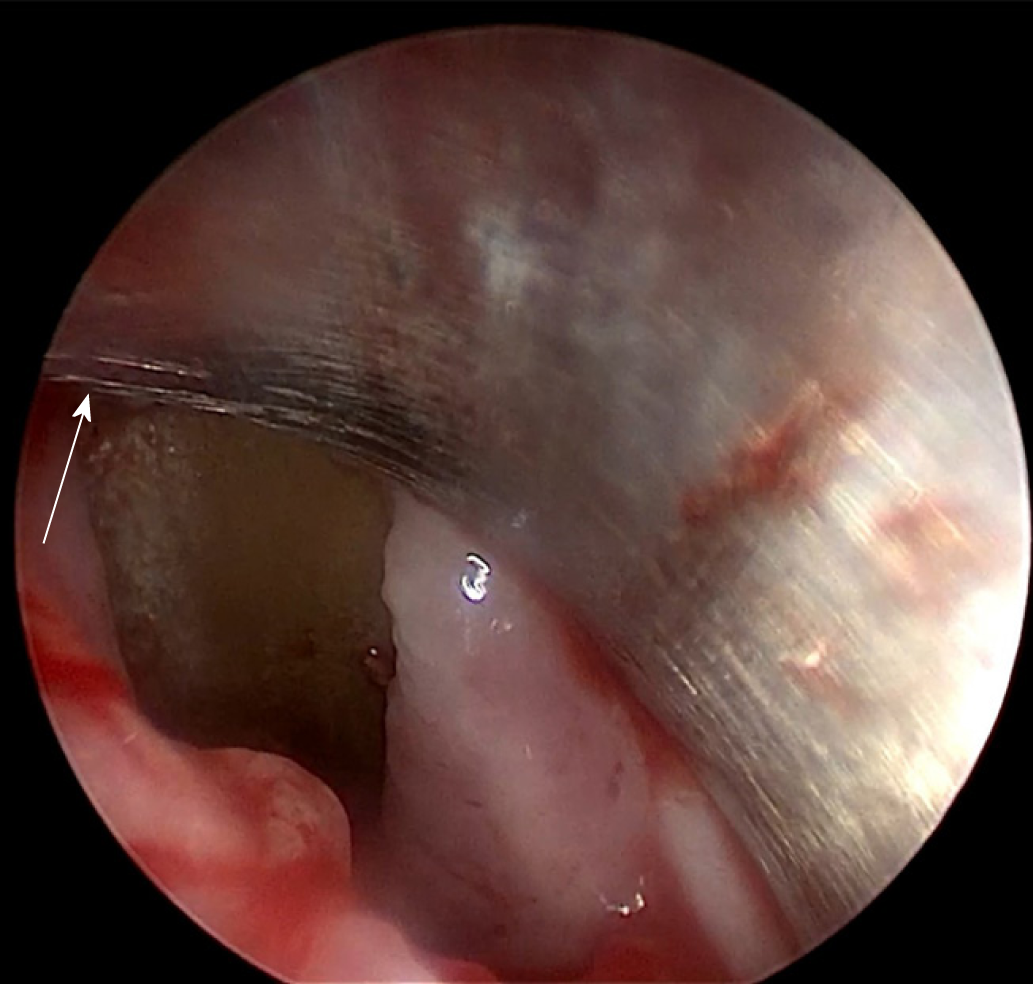
Figure 4 Endoscopic view of a black-brown foreign body penetrating the lateral wall of the esophagus, surrounding an edematous esophageal mucosa (Arrow: penetrated site).
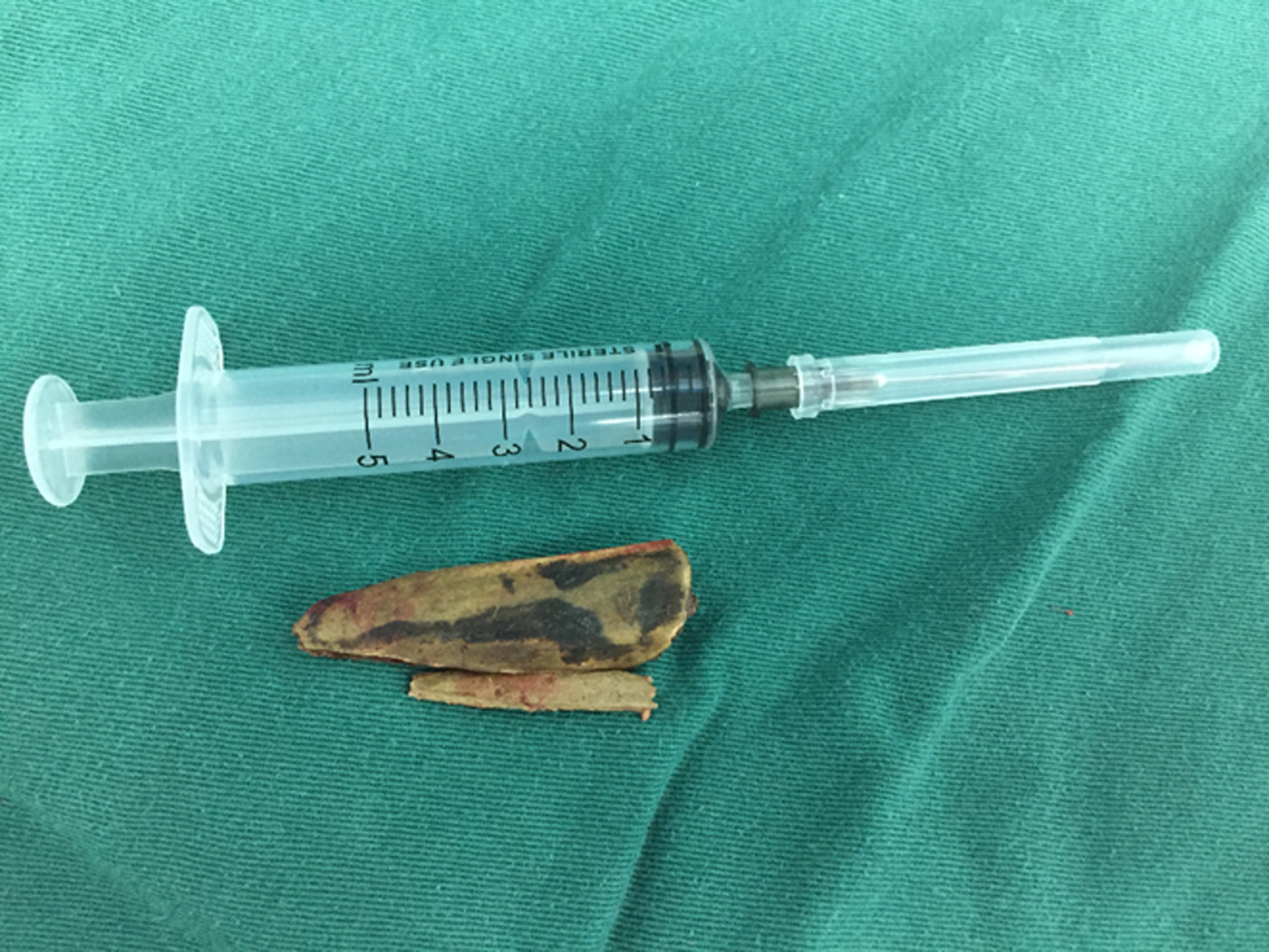
Figure 5 Complete removal of two foreign bodies.
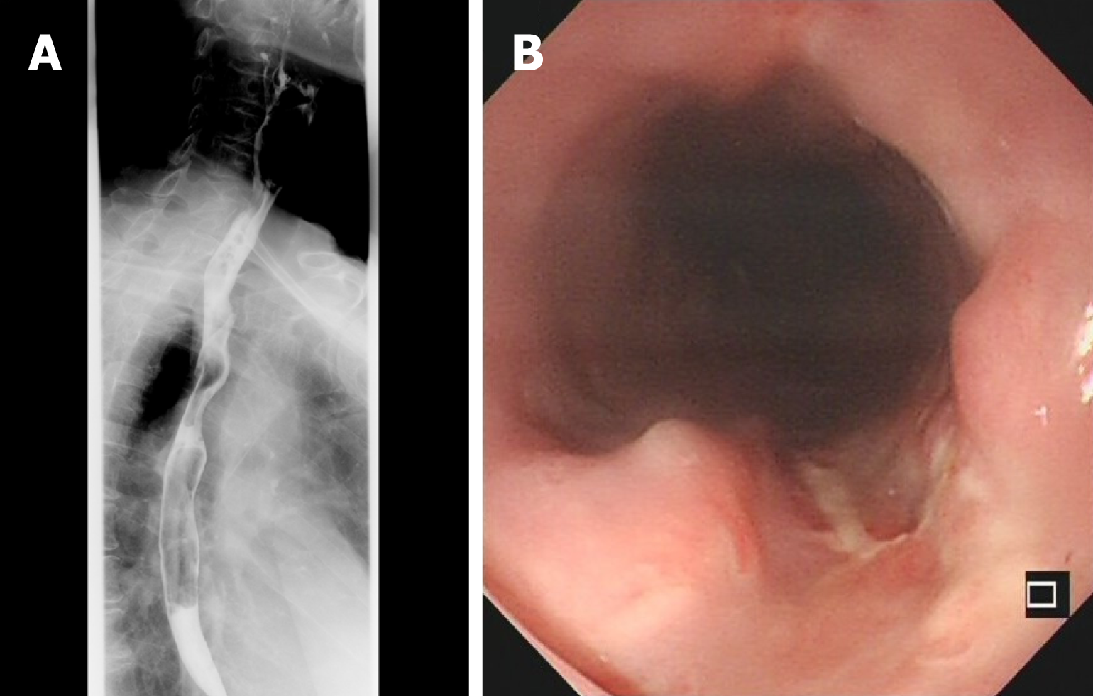
Figure 6 Upper gastrointestinal imaging on postoperative day 14.
A: Upper gastrointestinal enterography image showing no contrast agent leakage; B: Flexible endoscopy image showing a superficial esophageal ulcer without evident perforation 22–23 cm from the incisor.
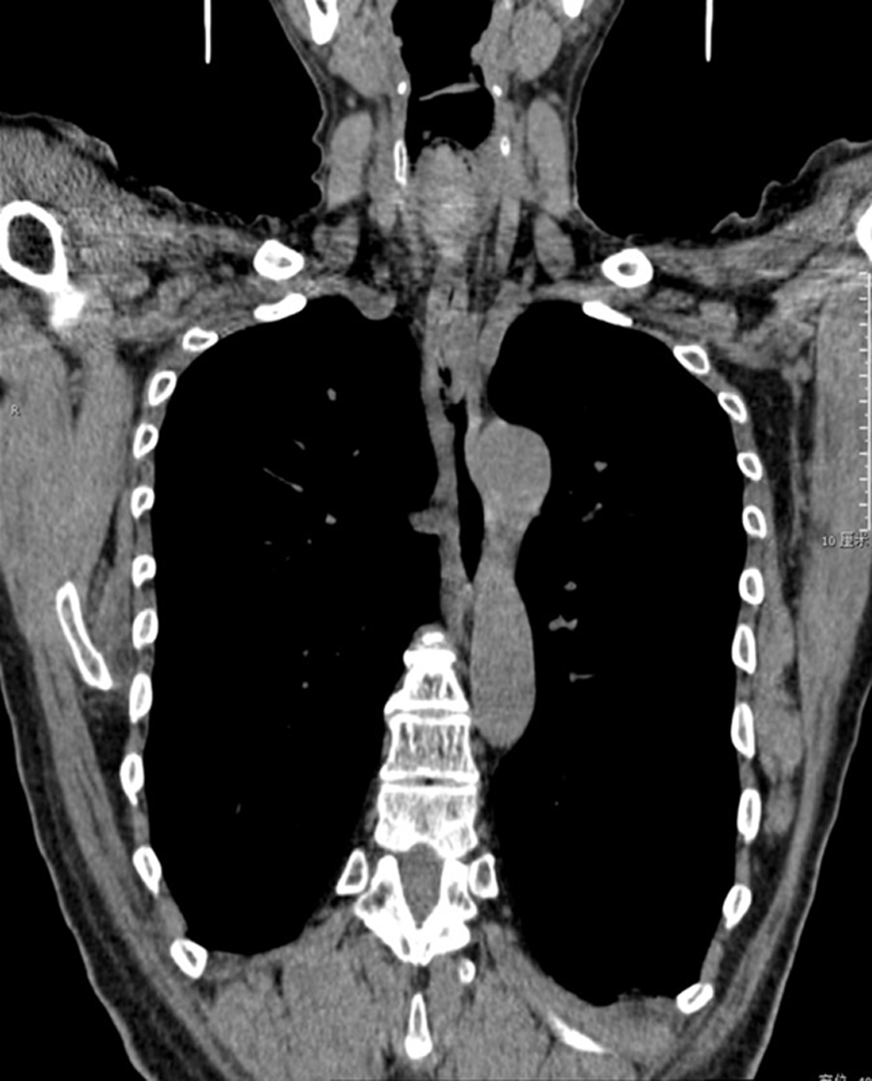
Figure 7 Chest coronal computed tomography image showing no residual foreign body or infection in the mediastinum after oral contrast.
- Citation: Wang D, Gao CB. Rigid esophagoscopy combined with angle endoscopy for treatment of superior mediastinal foreign bodies penetrating into the esophagus caused by neck trauma: A case report. World J Clin Cases 2019; 7(23): 4130-4136
- URL: https://www.wjgnet.com/2307-8960/full/v7/i23/4130.htm
- DOI: https://dx.doi.org/10.12998/wjcc.v7.i23.4130


