Copyright
©The Author(s) 2019.
World J Clin Casesol. Sep 26, 2019; 7(18): 2864-2870
Published online Sep 26, 2019. doi: 10.12998/wjcc.v7.i18.2864
Published online Sep 26, 2019. doi: 10.12998/wjcc.v7.i18.2864
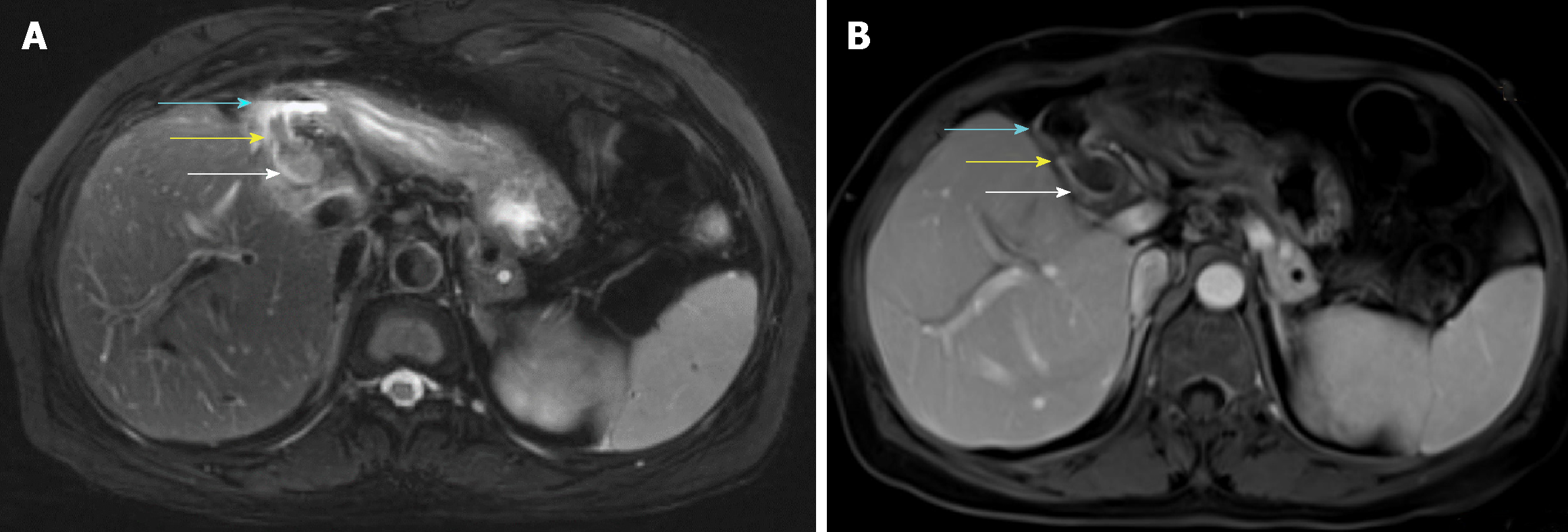
Figure 1 Magnetic resonance imaging.
A: Axial T2 weighted image showing a fistula connecting the bile duct (white arrow) and the duodenal bulb (blue arrow) which had a gas-liquid plane inside. Inside the fistula, there was a hypointense lesion (yellow arrow); B: Axial T1 weighted image contrast enhanced showing no enhancement of the bile duct (white arrow), duodenal bulb (blue arrow), or lesion (yellow arrow).
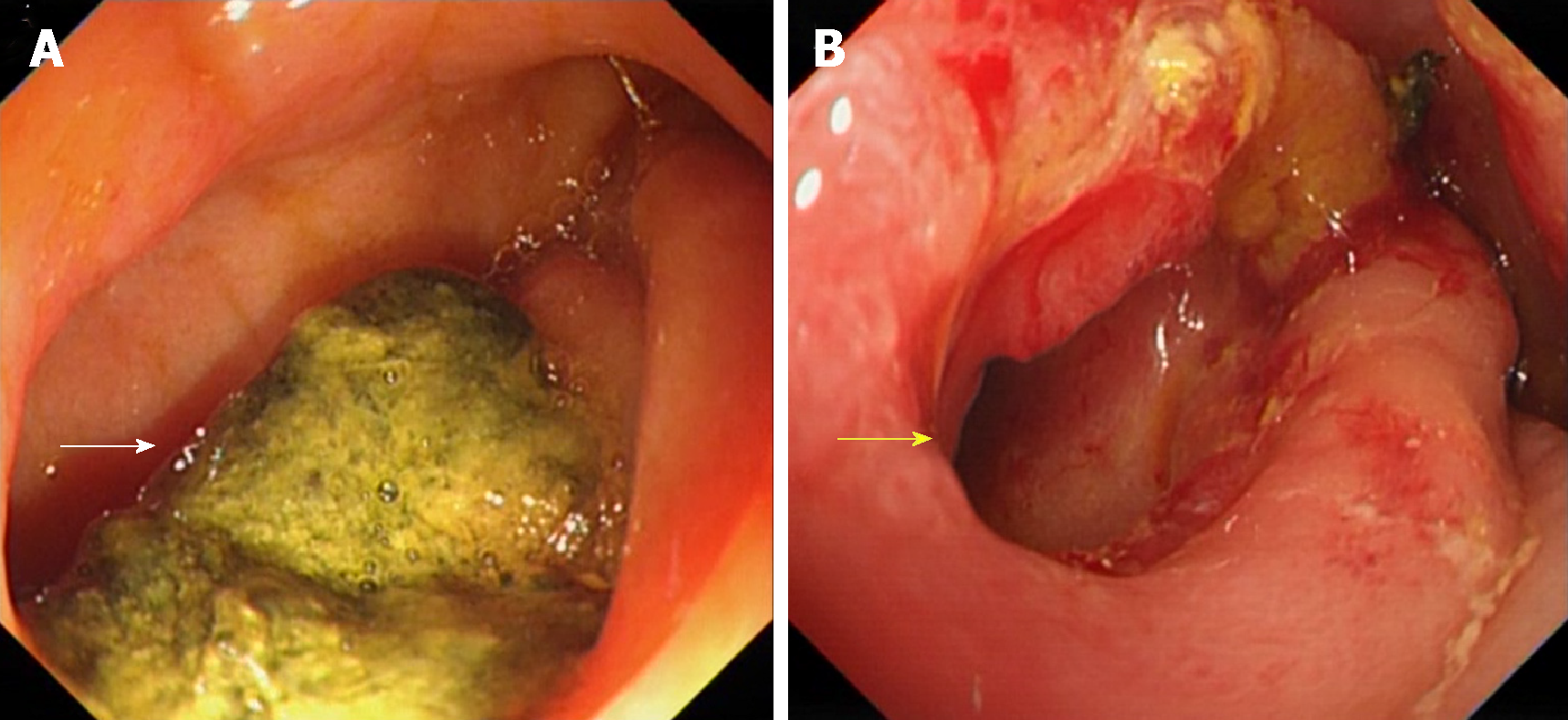
Figure 2 Gastroscopic images.
A: An incarcerated stone (White arrow); B: A fistula (yellow arrow) was observed in the duodenal bulb after the stone was removed.
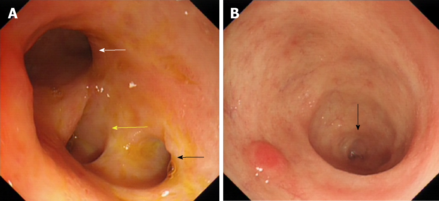
Figure 3 Gastroscopic images of the bile duct.
A: The image of hilar bile duct showing the left hepatic duct (white arrow), right anterior hepatic duct (yellow Arrow), and right posterior hepatic duct (black arrow); B: The terminal part (black arrow) of the common bile duct.
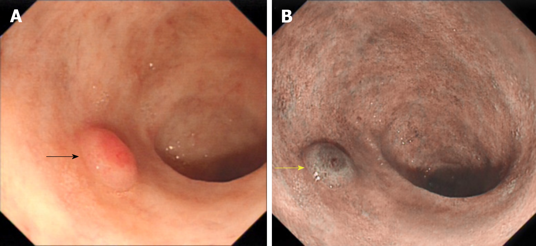
Figure 4 Gastroscopic images of the protruding lesion in the common bile duct.
A: The lesion under white light (black arrow); B: The lesion under narrow band imaging (yellow arrow).
Figure 5 Endoscopic mucosal resection of the lesion in the common bile duct.
A: The submucosal injection; B: The snare polypectomy of the polyp; C: Image collected after snare polypectomy.
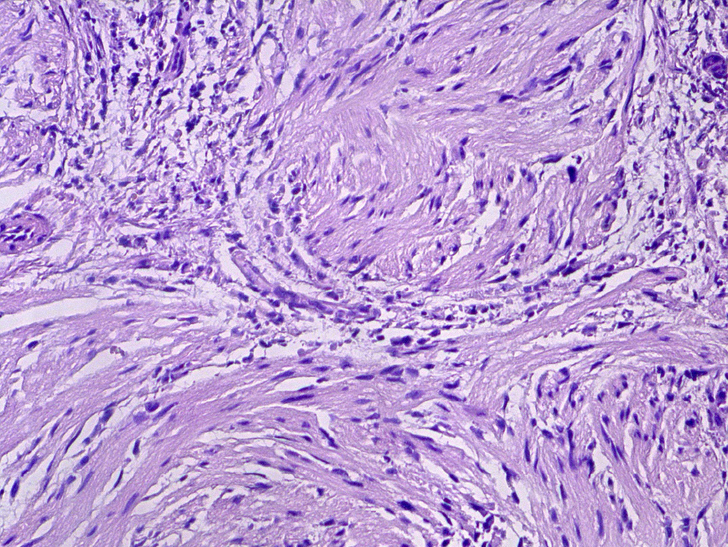
Figure 6 Histopathology.
Histological section showing an inflammatory fibroid polyp (hematoxylin and eosin staining; magnification, 200×).
- Citation: Yang S, Yang L, Wang XY, Yang YM. Endoscopic mucosal resection of a bile duct polyp: A case report. World J Clin Casesol 2019; 7(18): 2864-2870
- URL: https://www.wjgnet.com/2307-8960/full/v7/i18/2864.htm
- DOI: https://dx.doi.org/10.12998/wjcc.v7.i18.2864


