Copyright
©The Author(s) 2019.
World J Clin Cases. Sep 26, 2019; 7(18): 2658-2665
Published online Sep 26, 2019. doi: 10.12998/wjcc.v7.i18.2658
Published online Sep 26, 2019. doi: 10.12998/wjcc.v7.i18.2658
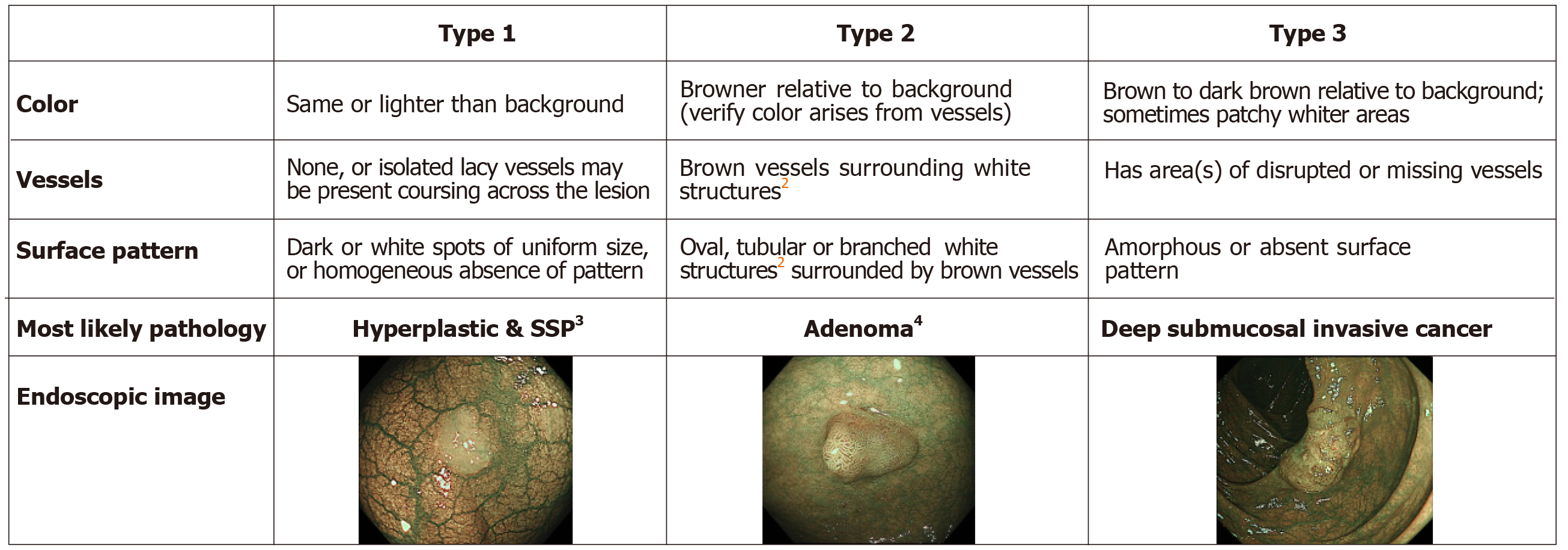
Figure 1 Narrow Band Imaging International Colorectal Endoscopic classification1.
1Can be applied using colonoscopes with/ without optical (zoom) magnification. 2These structures (regular or irregular) may represent the pits and the epithelium of the crypt opening. 3In the WHO classification, sessile serrated polyp and sessile serrated adenoma are synonymous. 4Type 2 consists of Vienna classification types 3, 4 and superficial 5 (all adenomas with either low or high grade dysplasia, or with superficial submucosal carcinoma). The presence of high grade dysplasia or superficial submucosal carcinoma may be suggested by an irregular vessel or surface pattern, and is often associated with atypical morphology. SSP: Sessile serrated polyp.
Figure 2 The Japan Narrow Band Imaging Expert Team classification.
1If visible, the caliber in the lesion is similar to surrounding normal mucosa. 2Micro-vessels are often distributed in a punctate pattern and well-ordered reticular or spiral vessels may not be observed in depressed lesions. 3Deep submucosal invasive cancer may be included.
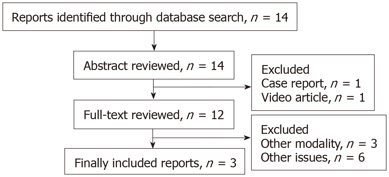
Figure 3 Flow diagram of study selection.
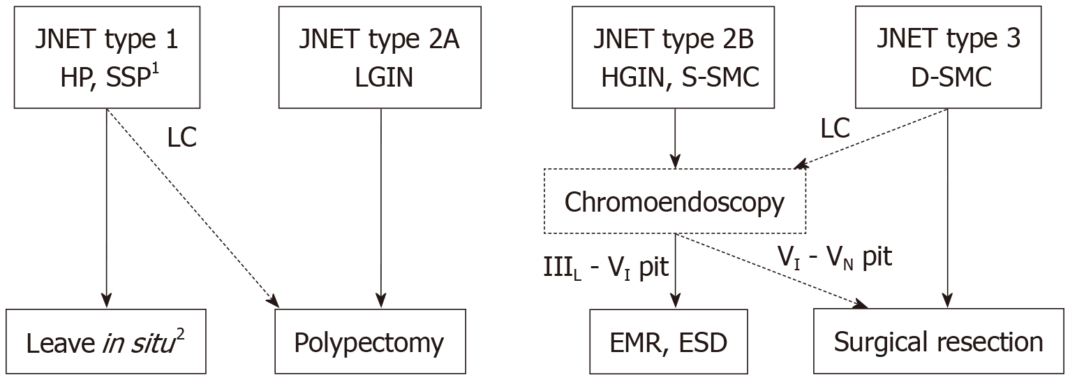
Figure 4 Treatment strategy of colorectal lesions using the Japan Narrow Band Imaging Expert Team classification.
1Any polyps suspected of SSPs should be resected by endoscopic treatment. 2Hyperplastic polyps less than 5mm in size are commonly left in situ. HP: Hyperplastic polyp; SSP: Sessile serrated polyp; LGIN: Low grade intramucosal neoplasia; HGIN: High grade intramucosal neoplasia; S-SMC: Shallow submucosal invasive cancer; D-SMC: Deep submucosal invasive cancer; LC: Low confidence.
- Citation: Hirata D, Kashida H, Iwatate M, Tochio T, Teramoto A, Sano Y, Kudo M. Effective use of the Japan Narrow Band Imaging Expert Team classification based on diagnostic performance and confidence level. World J Clin Cases 2019; 7(18): 2658-2665
- URL: https://www.wjgnet.com/2307-8960/full/v7/i18/2658.htm
- DOI: https://dx.doi.org/10.12998/wjcc.v7.i18.2658


