Copyright
©The Author(s) 2019.
World J Clin Cases. Sep 6, 2019; 7(17): 2573-2579
Published online Sep 6, 2019. doi: 10.12998/wjcc.v7.i17.2573
Published online Sep 6, 2019. doi: 10.12998/wjcc.v7.i17.2573
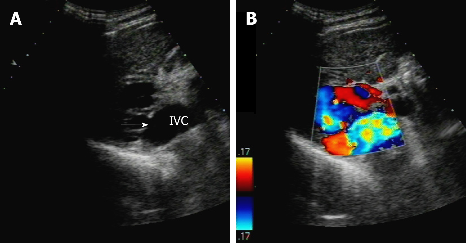
Figure 1 Ultrasonography revealed a spontaneous intrahepatic portosystemic shunt.
A: The branch of the right portal vein connects to the inferior vena cava through (arrow); B: The extrahepatic venous plexus showed turbulent spectrum locally. IVC: Inferior vena cava.
Figure 2 Abdomen enhanced computed tomography revealed a spontaneous intrahepatic portosystemic shunt.
A: The right branch of portal vein (PV) was thick and showed vermicular dilatation vein cluster (arrow); B: The left branch of PV was thin and occluded (arrow); C: The right branch of PV connected with inferior vena cava (arrowhead), extrahepatic dilatation and distortion of blood vessels (arrow).
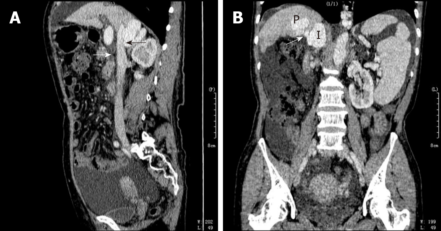
Figure 3 Abdomen enhanced computed tomography revealed a spontaneous intrahepatic portosystemic shunt at sagittal and coronal position.
A: The right branch of portal vein (PV) shunt out of the liver to communicate with inferior vena cava (IVC) (arrow) at sagittal position (black arrow); B: The right branch of PV shunt out of the liver to communicate with IVC (arrow) at coronal position.
- Citation: Tan YW, Sheng JH, Tan HY, Sun L, Yin YM. Rare spontaneous intrahepatic portosystemic shunt in hepatitis B-induced cirrhosis: A case report. World J Clin Cases 2019; 7(17): 2573-2579
- URL: https://www.wjgnet.com/2307-8960/full/v7/i17/2573.htm
- DOI: https://dx.doi.org/10.12998/wjcc.v7.i17.2573


