Copyright
©The Author(s) 2019.
World J Clin Cases. Sep 6, 2019; 7(17): 2562-2566
Published online Sep 6, 2019. doi: 10.12998/wjcc.v7.i17.2562
Published online Sep 6, 2019. doi: 10.12998/wjcc.v7.i17.2562
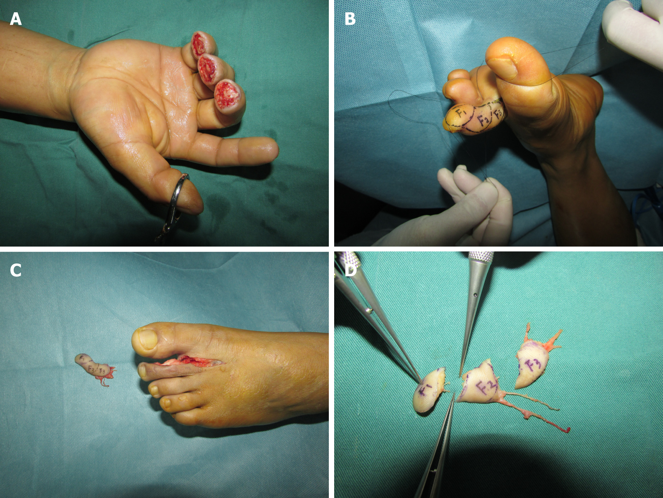
Figure 1 Split tibial second toe flap.
A: Three days after the first stage surgery, the wounds were clean and ready for free flap transfer; B: A 1.6 cm2 × 4.5 cm2 tibial second toe flap designed before operation; C: Harvested tibial second toe flap; D: Flap was split into three small flaps as F1, F2, and F3 under a microscope.
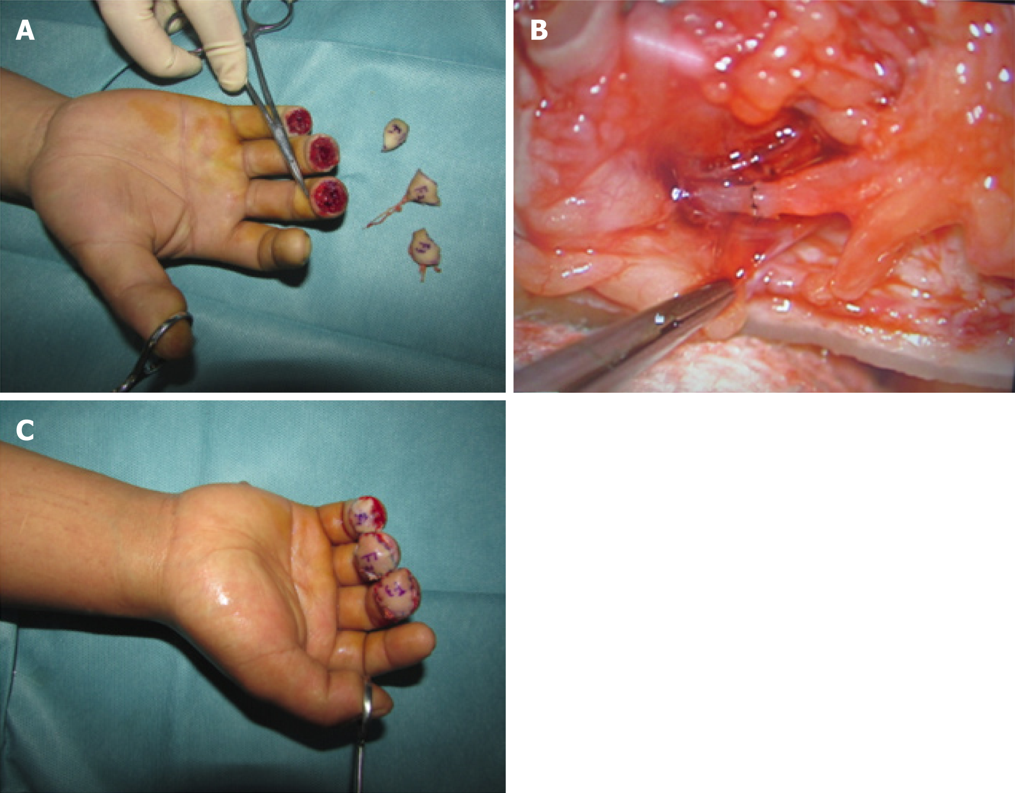
Figure 2 F3, F2, and F1 were transferred to middle, ring, and little fingertips with the one artery.
A: F1, F2, and F3 flaps before transfer to recipient sites; B: Representative anastomosis of the vessels and nerve; C: F1, F2, and F3 flaps transferred to recipient sites.
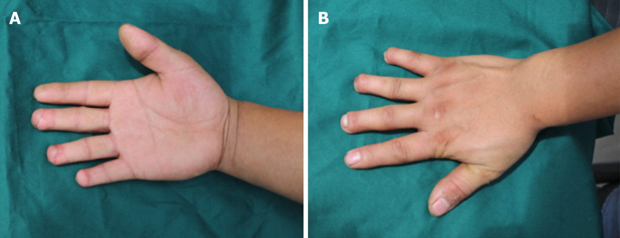
Figure 3 Six months after the surgery.
A: Palmar view; B: Dorsal view.
- Citation: Wang KL, Zhang ZQ, Buckwalter JA, Yang Y. Supermicrosurgery in fingertip defects-split tibial flap of the second toe to reconstruct multiple fingertip defects: A case report. World J Clin Cases 2019; 7(17): 2562-2566
- URL: https://www.wjgnet.com/2307-8960/full/v7/i17/2562.htm
- DOI: https://dx.doi.org/10.12998/wjcc.v7.i17.2562


