Copyright
©The Author(s) 2019.
World J Clin Cases. Aug 6, 2019; 7(15): 2087-2093
Published online Aug 6, 2019. doi: 10.12998/wjcc.v7.i15.2087
Published online Aug 6, 2019. doi: 10.12998/wjcc.v7.i15.2087
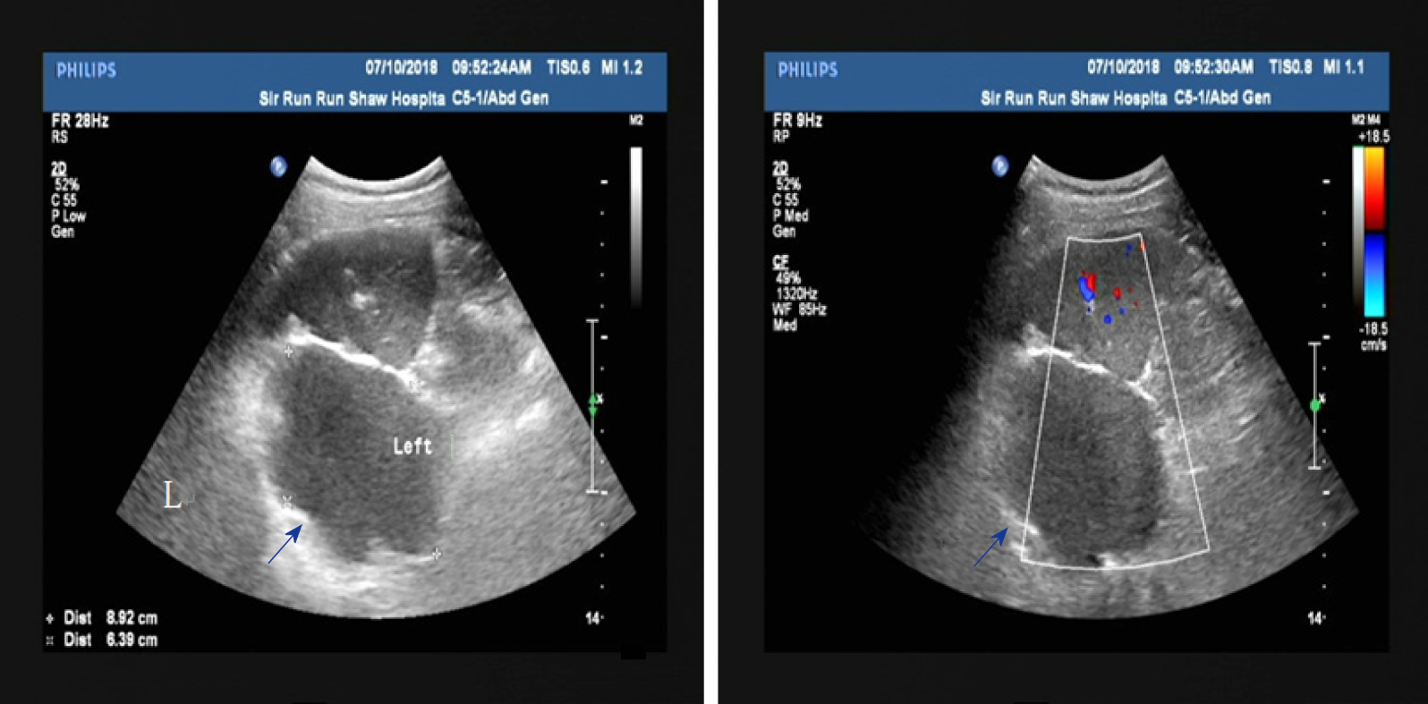
Figure 1 Ultrasound images of the gastric duplication cyst.
A large cystic focus (blue arrow) was visible in the space between the spleen, stomach, and left kidney, with no vascularization on Doppler.
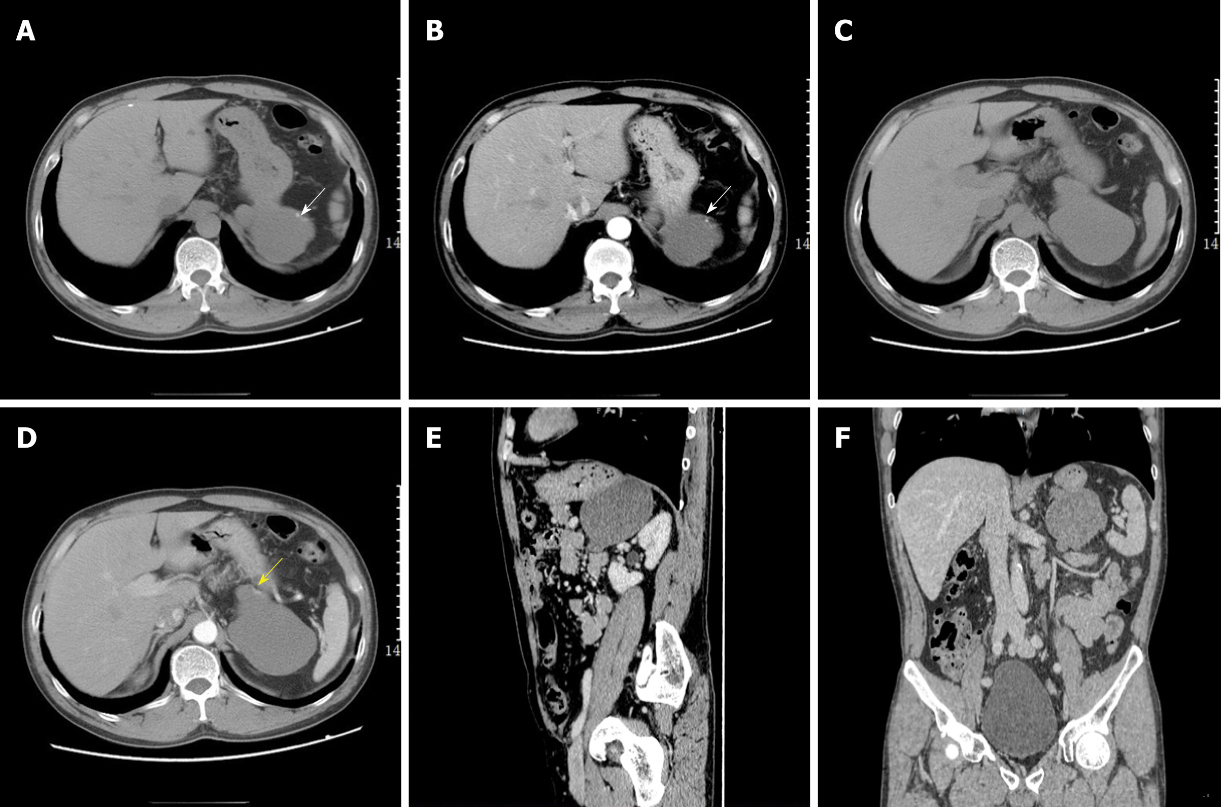
Figure 2 Contrast-enhanced computed tomography images of the gastric duplication cyst.
A-F: A large cystic hypodense focus was along the greater curvature of the stomach, measuring 95 mm × 61 mm × 66 mm; A and B: The focus was lobulated and well-circumscribed with a small amount of calcification on its wall (white arrow); C and D: A nutrient vessel from the stomach on the cyst wall was observed on arterial phase images (yellow arrow).
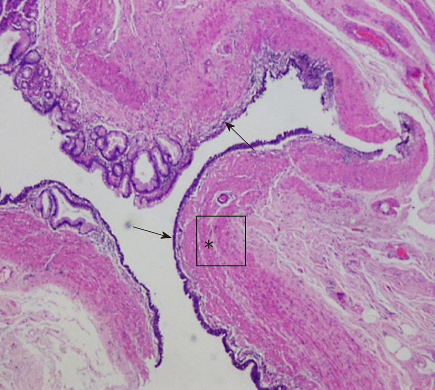
Figure 3 Photomicrograph (hematoxylin and eosin staining, original magnification, ×4) of the surgical specimen demonstrates an inner mucosal layer (arrows) within the cystic mass, along with a continuous muscular layer (asterisk) shared with the stomach.
- Citation: Xv FY, Sun A, Gan Y, Hu HJ. Gastric duplication cyst mimicking large cystic lymphangioma in an adult: A rare case report and review of the literature. World J Clin Cases 2019; 7(15): 2087-2093
- URL: https://www.wjgnet.com/2307-8960/full/v7/i15/2087.htm
- DOI: https://dx.doi.org/10.12998/wjcc.v7.i15.2087


