Copyright
©The Author(s) 2019.
World J Clin Cases. Aug 6, 2019; 7(15): 2049-2057
Published online Aug 6, 2019. doi: 10.12998/wjcc.v7.i15.2049
Published online Aug 6, 2019. doi: 10.12998/wjcc.v7.i15.2049
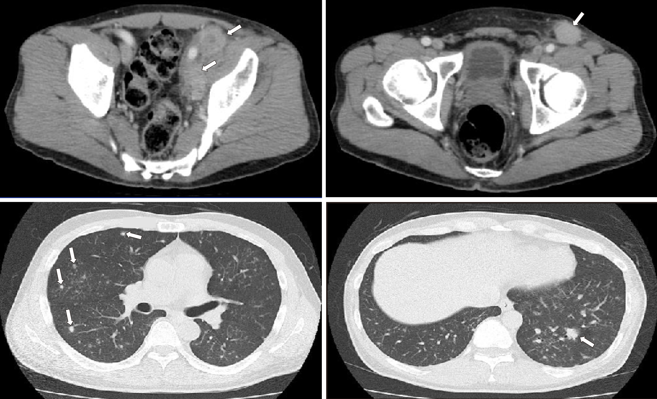
Figure 1 Computed tomography images.
(Upper row) Pelvic computed tomography scan demonstrated multiple lymph node lesions (arrows) continuing to the left common iliac artery (left)/left inguinal region (right). (Lower row) Scattered nodular lesions were found in both lung fields (arrows).
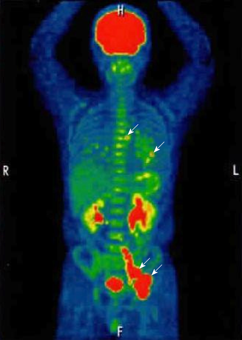
Figure 2 Positron emission tomography/computed tomography (longitudinal) image.
Arrows indicate lymphoma lesions. The mean of the standardized uptake value was 18.1 in the multiple lymph node lesions continuing to the mesentery and para-aortic/left common iliac artery/left inguinal, 3.5 in the lesions of the lung fields and the mediastinal nodes. No uptake in the stomach and duodenum.
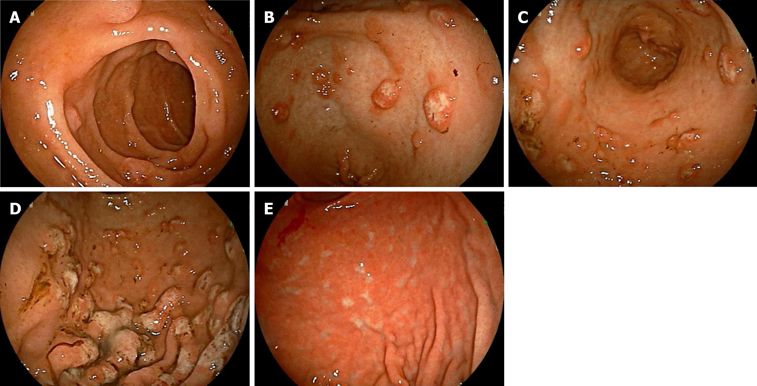
Figure 3 Esophagogastroduodenoscopy findings.
Multiple polypoid lesions of 2-3 mm in diameter were seen in the descending portion of the duodenum (A). Various large and small polypoid lesions were seen in the antrum of the stomach (B; lesser curvature side, C; extensive curvature side), accompanied by a change in white tone at the center. Mucosal folds in the corpus of the stomach at the extensive curvature side were slightly thickened, and its surface had changed to a white tone (D). After treatment, numerous white scars were found, and the thickened mucosal folds were improved in the stomach (E).
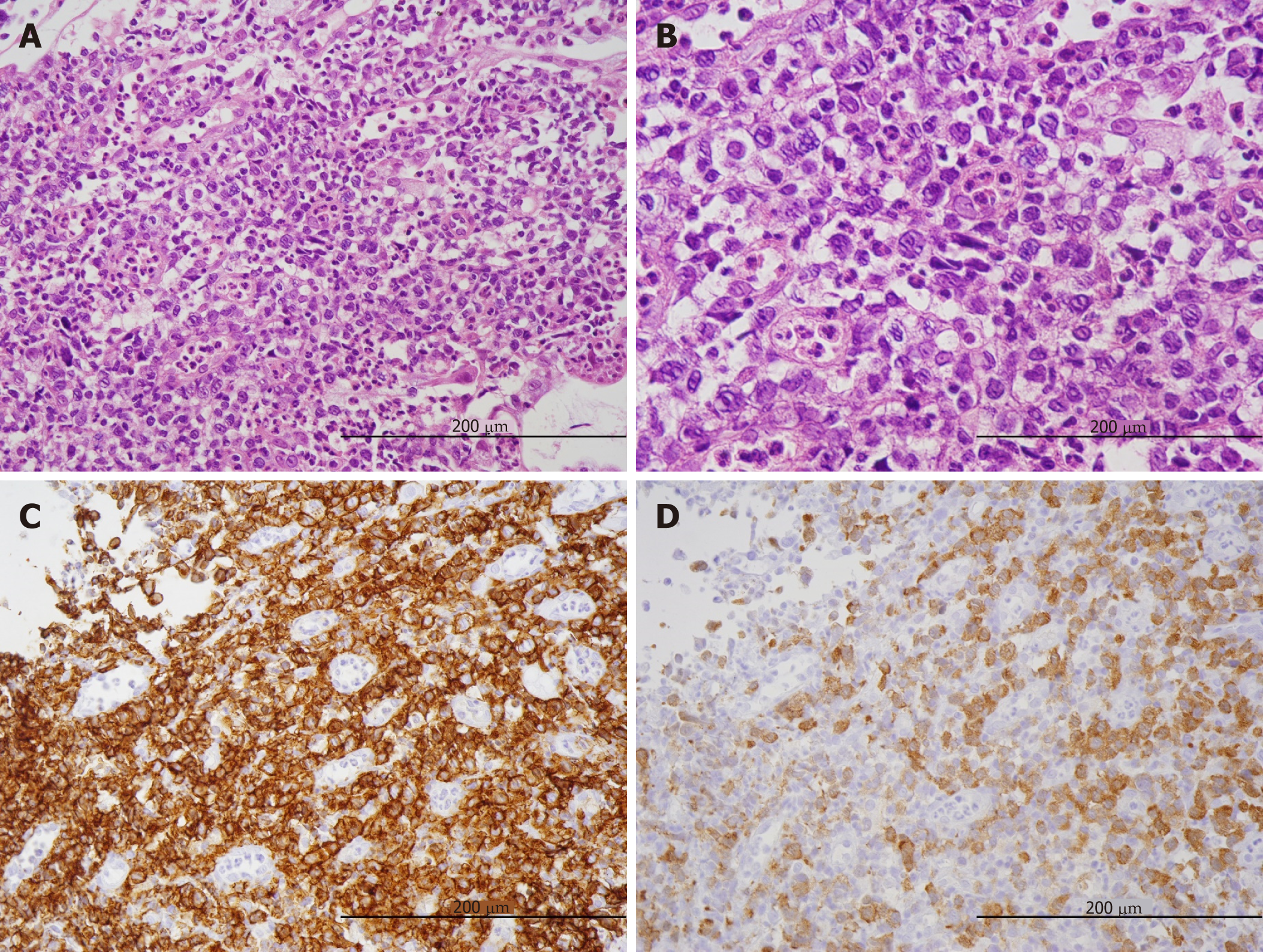
Figure 4 Histopathological findings in biopsy samples (x 200), Medium to large-sized abnormal lymphoid cells with irregular nuclei grew diffusely (A), and mitotic figures were observed in high numbers (B).
In immunostaining of lymphoma cells, CD30 was strongly positive (C), and ALK showed positive in nuclear and cytoplasmic patterns (D).
- Citation: Saito M, Izumiyama K, Ogasawara R, Mori A, Kondo T, Tanaka M, Morioka M, Miyashita K, Tanino M. ALK-positive anaplastic large cell lymphoma presenting multiple lymphomatous polyposis: A case report and literature review. World J Clin Cases 2019; 7(15): 2049-2057
- URL: https://www.wjgnet.com/2307-8960/full/v7/i15/2049.htm
- DOI: https://dx.doi.org/10.12998/wjcc.v7.i15.2049


