Copyright
©The Author(s) 2019.
World J Clin Cases. Jul 26, 2019; 7(14): 1844-1849
Published online Jul 26, 2019. doi: 10.12998/wjcc.v7.i14.1844
Published online Jul 26, 2019. doi: 10.12998/wjcc.v7.i14.1844
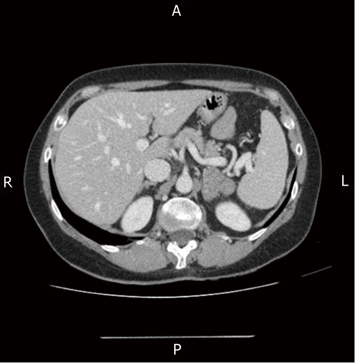
Figure 1 Computed tomography scan of abdomen showing left adrenal mass measuring 40 mm × 34 mm.
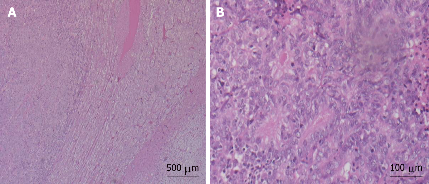
Figure 2 Hematoxylin and eosin stain of endometrial endometrioid carcinoma.
A: Panoramic view of left adrenal gland; B: High magnification showing the glandular pattern of neoplastic cells.
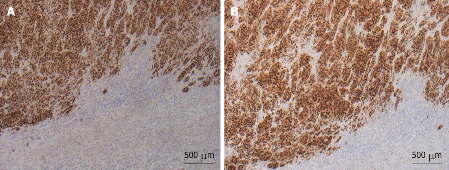
Figure 3 Immunohistochemical staining.
A, B: Immunohistochemical staining positive for estrogen receptors (A) And progesterone receptors (B) Shows high reactivity in metastatic cells.
- Citation: Da Dalt G, Friziero A, Grego A, Serafini S, Fassina A, Blandamura S, Sperti C. Adrenal metastasis from endometrial cancer: A case report. World J Clin Cases 2019; 7(14): 1844-1849
- URL: https://www.wjgnet.com/2307-8960/full/v7/i14/1844.htm
- DOI: https://dx.doi.org/10.12998/wjcc.v7.i14.1844


