Copyright
©The Author(s) 2019.
World J Clin Cases. Jun 6, 2019; 7(11): 1351-1357
Published online Jun 6, 2019. doi: 10.12998/wjcc.v7.i11.1351
Published online Jun 6, 2019. doi: 10.12998/wjcc.v7.i11.1351
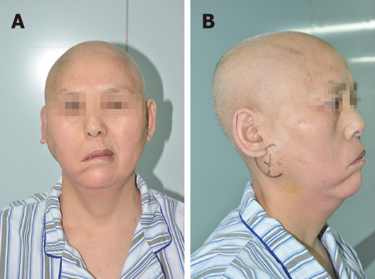
Figure 1 Clinical photographs of the right parotid swelling and characteristics of right facial palsy.
A: Front photograph; B: Right side photograph. (March 2017)
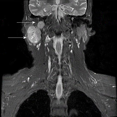
Figure 2 Pre-operative magnetic resonance imaging of the tumour (coronal, April 2017).
A 3.9 cm × 2.9 cm × 5.3 cm sized lobulated, well-encapsulated, and heterogeneously enhancing mass on the deep lobe of the right parotid gland (arrow). It extended into the mastoid and was associated with bone destruction in the wall of the ear canal and mastoid.
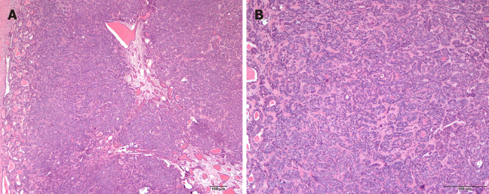
Figure 3 Photomicrographs of the trabecular type of basal cell adenoma.
A: A capsule and an eosinophilic basal membrane-like structure (hematoxylin-eosin staining, × 40); B: Small cuboidal to columnar epithelial cells arranged in strands with round hyperchromatic nuclei and relatively rounded central cells. The strands are surrounded by hyalinised connective tissue (hematoxylin-eosin staining, × 100).
Figure 4 Intra-operative appearance of the tumour mass.
A: The right parotid gland part of the tumour was separated; B: The basicranial part of the tumour was removed; C: The right mastoid process and cerebellar dura mater were resected. The mass was clearly lobular in shape, and the mastoid was partially destroyed. (April 2017)
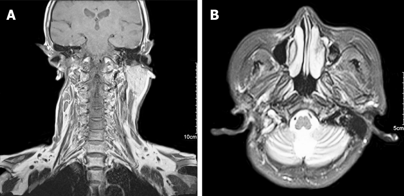
Figure 5 Post-operative magnetic resonance imaging of the patient 3 mo after operation.
No obvious residual tissue and no recurrence of the tumour. A: Coronal; B: Axial. (July 2017)
- Citation: Du LY, Weng XH, Shen ZY, Cheng B. A large basal cell adenoma extending to the ipsilateral skull base and mastoid in the right parotid gland: A case report. World J Clin Cases 2019; 7(11): 1351-1357
- URL: https://www.wjgnet.com/2307-8960/full/v7/i11/1351.htm
- DOI: https://dx.doi.org/10.12998/wjcc.v7.i11.1351


