Copyright
©The Author(s) 2019.
World J Clin Cases. Jun 6, 2019; 7(11): 1351-1357
Published online Jun 6, 2019. doi: 10.12998/wjcc.v7.i11.1351
Published online Jun 6, 2019. doi: 10.12998/wjcc.v7.i11.1351
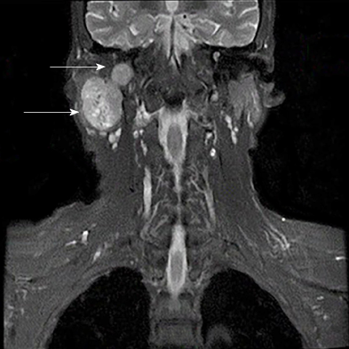
Figure 2 Pre-operative magnetic resonance imaging of the tumour (coronal, April 2017).
A 3.9 cm × 2.9 cm × 5.3 cm sized lobulated, well-encapsulated, and heterogeneously enhancing mass on the deep lobe of the right parotid gland (arrow). It extended into the mastoid and was associated with bone destruction in the wall of the ear canal and mastoid.
- Citation: Du LY, Weng XH, Shen ZY, Cheng B. A large basal cell adenoma extending to the ipsilateral skull base and mastoid in the right parotid gland: A case report. World J Clin Cases 2019; 7(11): 1351-1357
- URL: https://www.wjgnet.com/2307-8960/full/v7/i11/1351.htm
- DOI: https://dx.doi.org/10.12998/wjcc.v7.i11.1351


