Copyright
©The Author(s) 2019.
World J Clin Cases. Jan 6, 2019; 7(1): 73-78
Published online Jan 6, 2019. doi: 10.12998/wjcc.v7.i1.73
Published online Jan 6, 2019. doi: 10.12998/wjcc.v7.i1.73
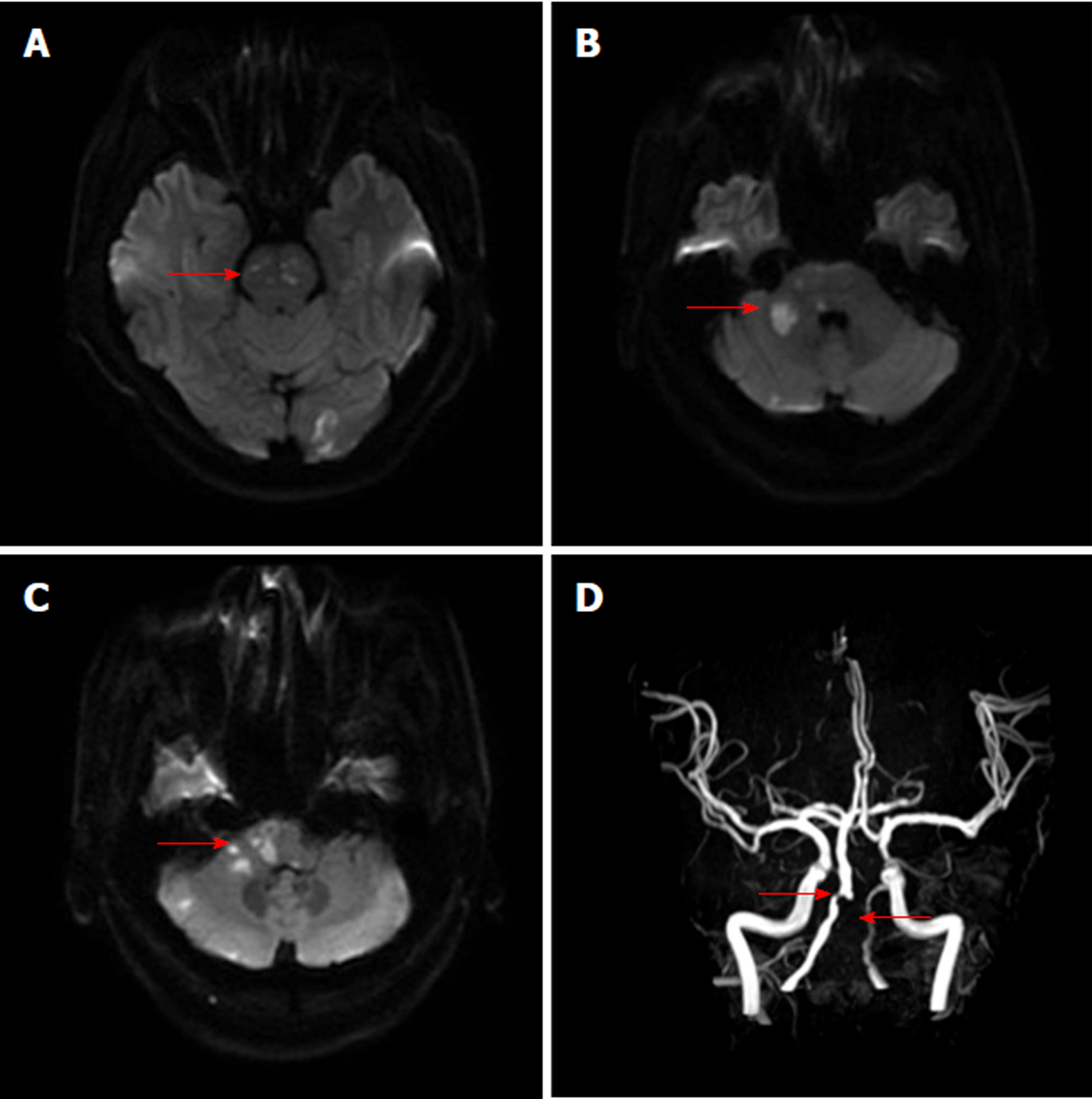
Figure 1 Brain magnetic resonance imaging and magnetic resonance angiography show multiple infarctions and occlusion and stenosis of the vertebral artery.
A-C: Diffusion weighted imaging shows acute multifocal infarctions in the pons, ventral medulla oblongata, cerebellopontine angle, and left occipital lobe; D: Brain magnetic resonance angiography indicates the occlusion of the left vertebral artery and severe stenosis of the proximal right vertebral artery.
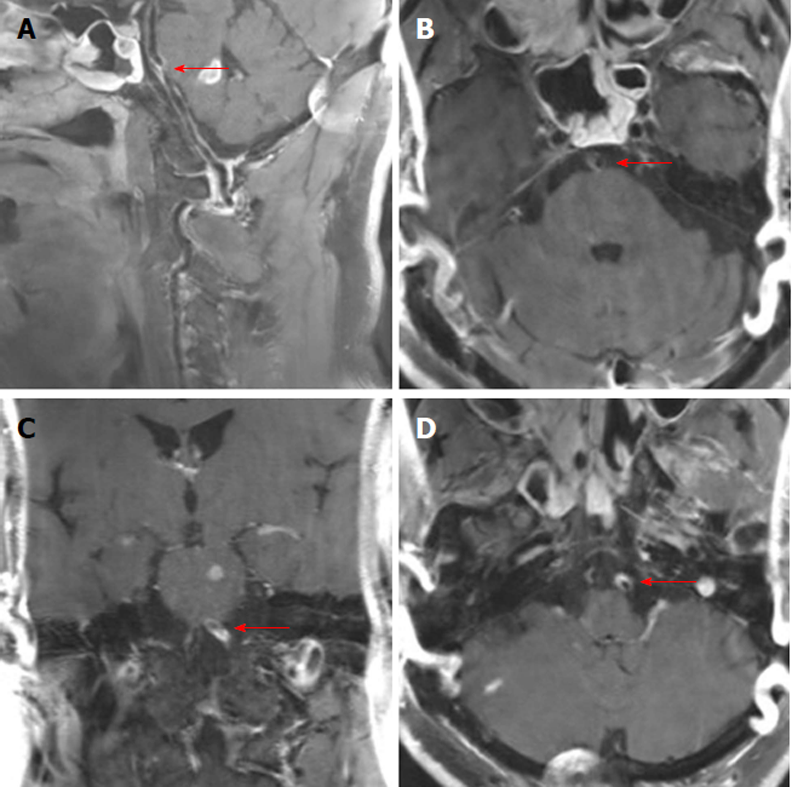
Figure 2 High-resolution magnetic resonance imaging shows the dissection of the basilar artery and left vertebral artery.
A, B: The eccentric periluminal hematoma of the basilar artery; C, D: The eccentric periluminal hematoma of the left vertebral artery.
- Citation: Li XT, Yuan JL, Hu WL. Vertebrobasilar artery dissection manifesting as Millard-Gubler syndrome in a young ischemic stroke patient: A case report. World J Clin Cases 2019; 7(1): 73-78
- URL: https://www.wjgnet.com/2307-8960/full/v7/i1/73.htm
- DOI: https://dx.doi.org/10.12998/wjcc.v7.i1.73


