Copyright
©The Author(s) 2019.
World J Clin Cases. Jan 6, 2019; 7(1): 49-57
Published online Jan 6, 2019. doi: 10.12998/wjcc.v7.i1.49
Published online Jan 6, 2019. doi: 10.12998/wjcc.v7.i1.49
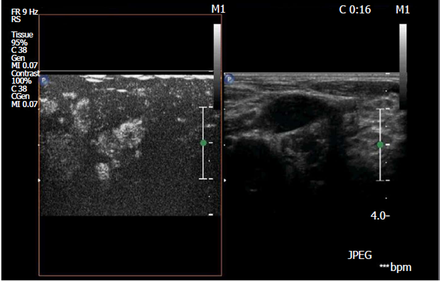
Figure 1 Ultrasound contrast sonogram of metastatic lymph nodes.
It reveals that the lymph nodes were enhanced, and the distribution was not uniform.
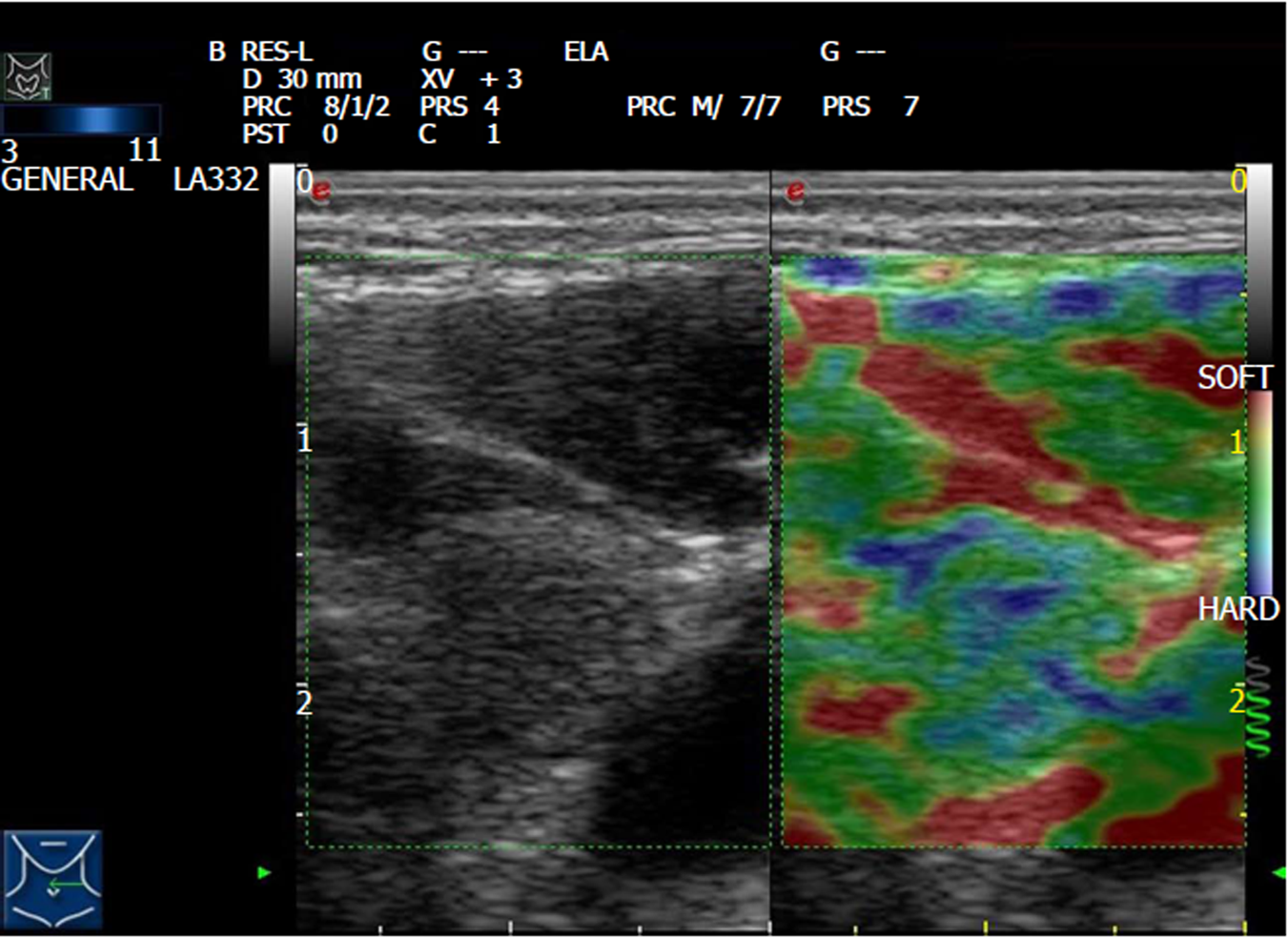
Figure 2 Elastography of metastatic lymph nodes in papillary thyroid carcinoma.
It reveals that the lymph nodes had a hard texture and blue-green color.
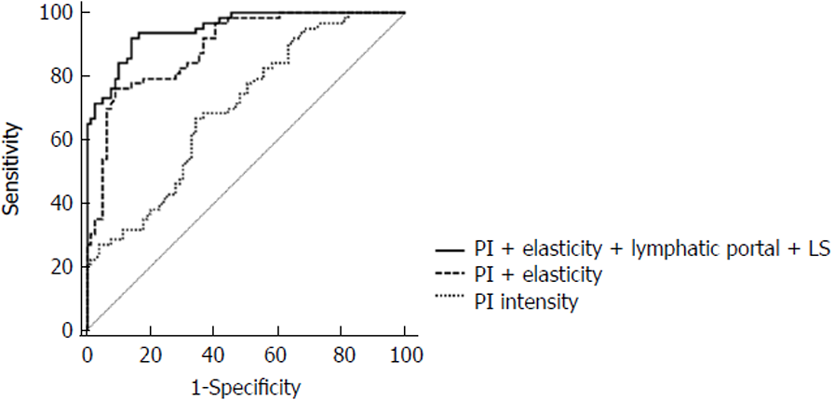
Figure 3 Receiver operating characteristic curve analysis of combined evaluation of papillary thyroid carcinoma cervical lymph node metastasis.
- Citation: Jiang W, Wei HY, Zhang HY, Zhuo QL. Value of contrast-enhanced ultrasound combined with elastography in evaluating cervical lymph node metastasis in papillary thyroid carcinoma. World J Clin Cases 2019; 7(1): 49-57
- URL: https://www.wjgnet.com/2307-8960/full/v7/i1/49.htm
- DOI: https://dx.doi.org/10.12998/wjcc.v7.i1.49


