Copyright
©The Author(s) 2019.
World J Clin Cases. Jan 6, 2019; 7(1): 116-121
Published online Jan 6, 2019. doi: 10.12998/wjcc.v7.i1.116
Published online Jan 6, 2019. doi: 10.12998/wjcc.v7.i1.116
Figure 1 Computed tomography scans of the tumor.
Figure 2 Gross anatomy of the spited tumor and its histology.
A: Gross anatomy of tumor, spited specimens; B: Histology by H and E staining shows the mild shapes of follicular dendritic cell sarcoma, comprised of fascicular oval and spindle cells, as well as infiltration of small lymphocytes into the tumor (200 × magnification); C: The high power field of histology by H and E staining shows that cytoplasm is abundant and eosinophilic. The cell nuclei are small and clear with ovalular or long spindle shapes (400 × magnification).
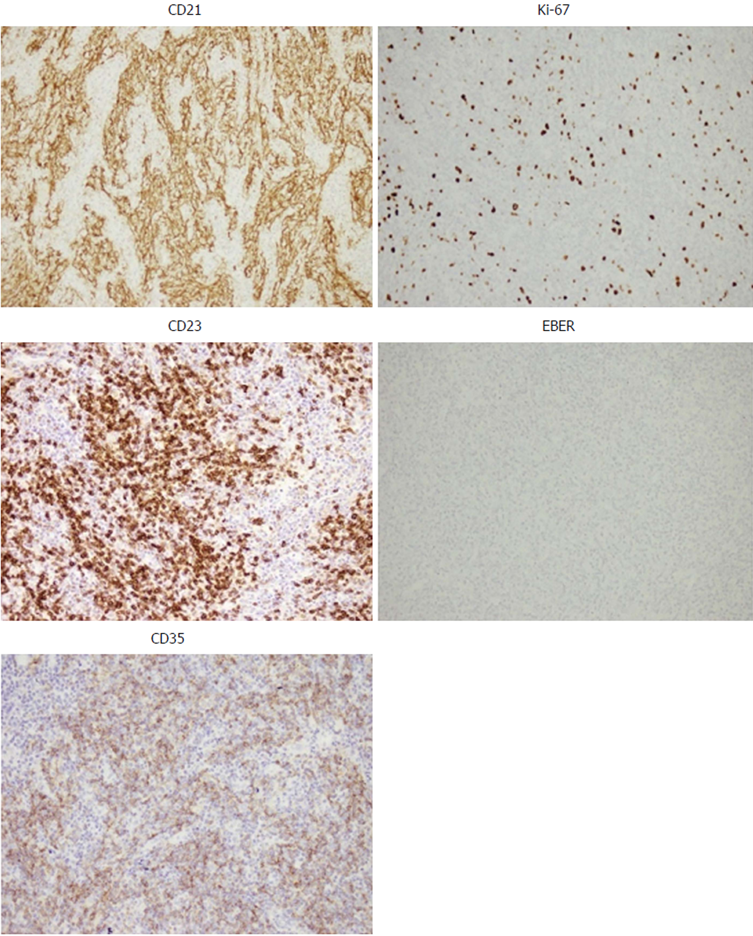
Figure 3 Immunohistochemical staining of tumor tissues.
Immunohistochemical images show positive staining for CD21, CD23 and CD35, which are specific biomarkers for diagnosing follicular dendritic cell sarcoma. Ki-67 staining shows 20% positive cells, while Epstein-Barr-encoded RNA was used as a negative stain. 200 × magnification for all images.
- Citation: Yan WX, Yu YX, Zhang P, Liu XK, Li Y. Follicular dendritic cell sarcoma detected in hepatogastric ligament: A case report and review of the literature. World J Clin Cases 2019; 7(1): 116-121
- URL: https://www.wjgnet.com/2307-8960/full/v7/i1/116.htm
- DOI: https://dx.doi.org/10.12998/wjcc.v7.i1.116


