Copyright
©The Author(s) 2018.
World J Clin Cases. Dec 26, 2018; 6(16): 1199-1201
Published online Dec 26, 2018. doi: 10.12998/wjcc.v6.i16.1199
Published online Dec 26, 2018. doi: 10.12998/wjcc.v6.i16.1199
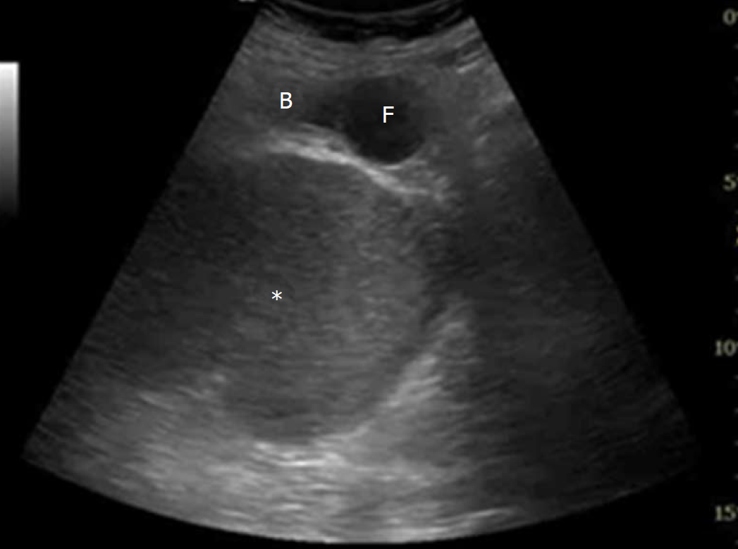
Figure 1 Bladder ultrasonography revealed a pelvic cystic lesion (asterisk) with mass effect on the bladder.
B: Bladder; F: Foley.
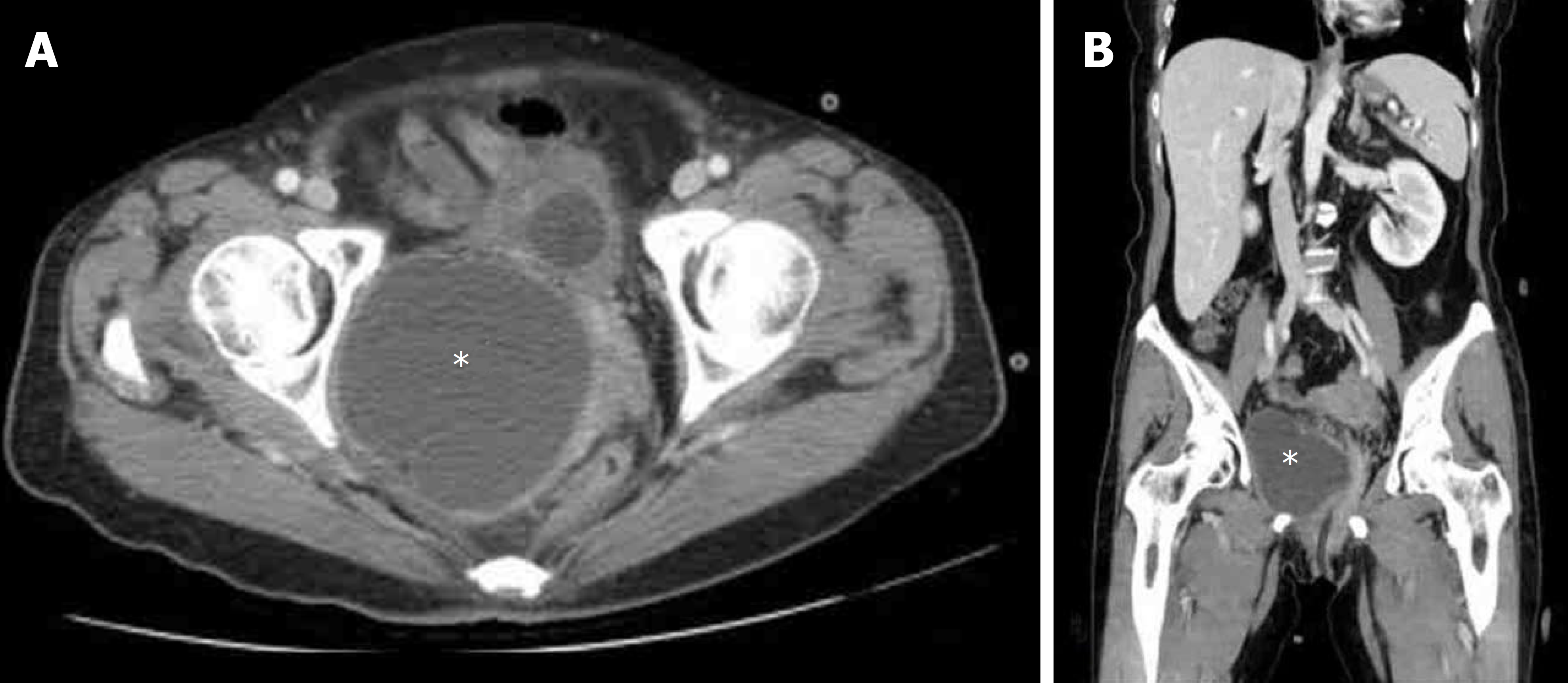
Figure 2 Pelvic computed tomography with axial (A) and sagittal (B) view showed a 9.
1 cm cystic lesion (asterisks) with homogeneous content in right lower pelvic region.
- Citation: Yeh CC, Yang SSD, Huang SC, Wang YC. Uncommon cause of voiding dysfunction in a female patient-vaginal abscess: A case report. World J Clin Cases 2018; 6(16): 1199-1201
- URL: https://www.wjgnet.com/2307-8960/full/v6/i16/1199.htm
- DOI: https://dx.doi.org/10.12998/wjcc.v6.i16.1199


