Copyright
©The Author(s) 2018.
World J Clin Cases. Dec 26, 2018; 6(16): 1175-1181
Published online Dec 26, 2018. doi: 10.12998/wjcc.v6.i16.1175
Published online Dec 26, 2018. doi: 10.12998/wjcc.v6.i16.1175
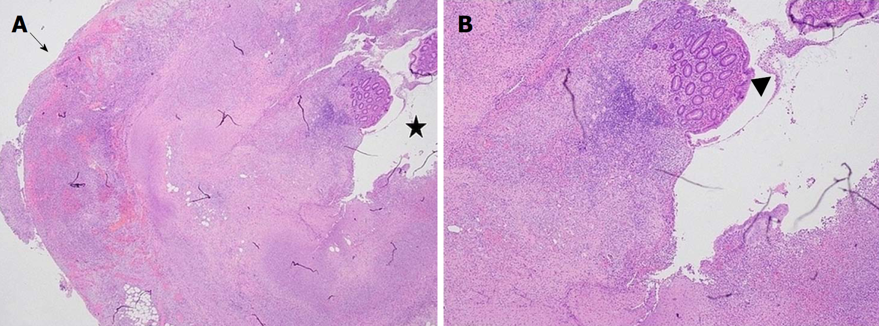
Figure 1 Histopathology of appendiceal specimen with appendicitis.
A: This slide is a cross-section of the appendix showing the appendiceal lumen indicated by the star displaying extensive ulceration and pervasive inflammation; with minimal residual mucosa. The transmural inflammation, involves the serosa as shown by the arrow; and depicts mucosal ulceration and necrosis; B: This slide image, photographed at a higher magnification, shows the residual mucosa (which appears normal) surrounded by necrotic tissue and cellular debris. The residual mucosa is indicated by the arrowhead.
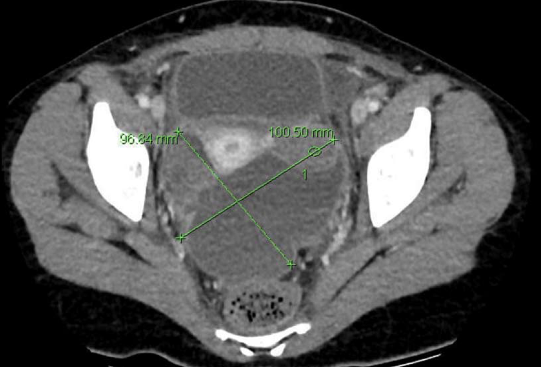
Figure 2 Pelvic collection - axial view.
The computerised tomography (CT) scan on postoperative day 4 shows a large pelvic collection with dimensions of 10.1 cm × 9.6 cm. This CT view is the axial view. The collection shows different segments displaying different densities.
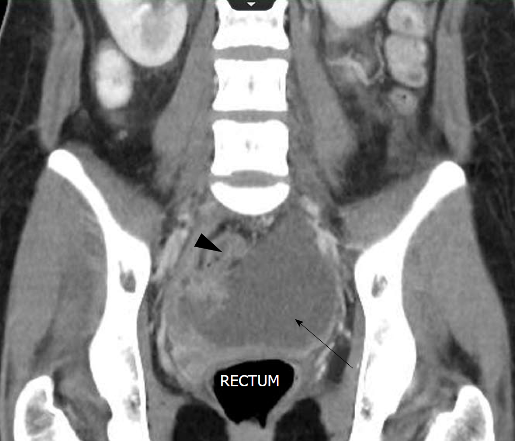
Figure 3 Pelvic collection - coronal view.
The computerised tomography (CT) scan on postoperative day 4 shows a large pelvic collection with dimensions of 10.1 cm × 9.6 cm, as indicated by the arrow. This CT view is the coronal view. The pelvic collection displays different segments portraying different densities, as well as loculated portions as shown by the arrowhead.
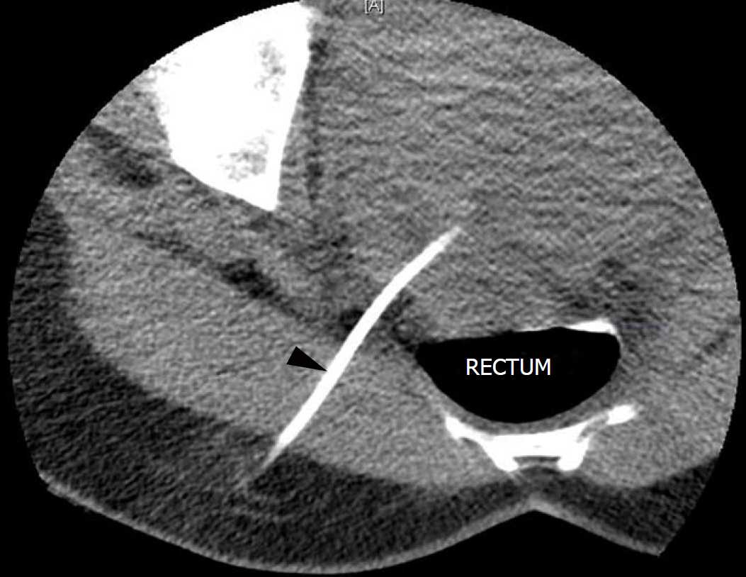
Figure 4 Percutaneous drainage of pelvic collection - axial view.
The large pelvic collection (10.1 cm × 9.6 cm) was drained percutaneously under computerised tomography guidance, and an 8 French Pigtail drain was left in situ in the rectouterine pouch of Douglas, as evident by the radio-opaque linear object at the 7 o’clock position, as indicated by the arrowhead.
- Citation: Tse A, Cheluvappa R, Selvendran S. Post-appendectomy pelvic abscess with extended-spectrum beta-lactamase producing Escherichia coli: A case report and review of literature. World J Clin Cases 2018; 6(16): 1175-1181
- URL: https://www.wjgnet.com/2307-8960/full/v6/i16/1175.htm
- DOI: https://dx.doi.org/10.12998/wjcc.v6.i16.1175


