
















©The Author(s) 2018.
World J Clin Cases. Dec 26, 2018; 6(16): 1175-1181
Published online Dec 26, 2018. doi: 10.12998/wjcc.v6.i16.1175
Published online Dec 26, 2018. doi: 10.12998/wjcc.v6.i16.1175
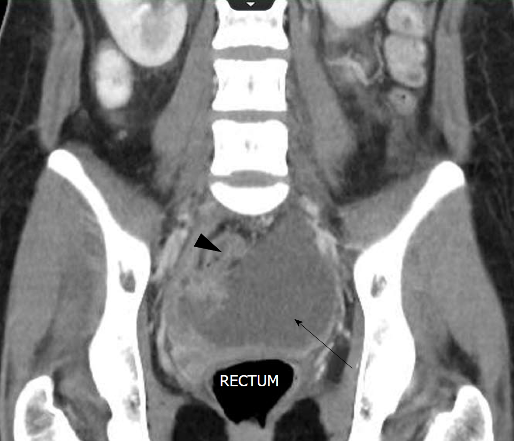
Figure 3 Pelvic collection - coronal view.
The computerised tomography (CT) scan on postoperative day 4 shows a large pelvic collection with dimensions of 10.1 cm × 9.6 cm, as indicated by the arrow. This CT view is the coronal view. The pelvic collection displays different segments portraying different densities, as well as loculated portions as shown by the arrowhead.
- Citation: Tse A, Cheluvappa R, Selvendran S. Post-appendectomy pelvic abscess with extended-spectrum beta-lactamase producing Escherichia coli: A case report and review of literature. World J Clin Cases 2018; 6(16): 1175-1181
- URL: https://www.wjgnet.com/2307-8960/full/v6/i16/1175.htm
- DOI: https://dx.doi.org/10.12998/wjcc.v6.i16.1175