Copyright
©The Author(s) 2018.
World J Clin Cases. Dec 6, 2018; 6(15): 1047-1052
Published online Dec 6, 2018. doi: 10.12998/wjcc.v6.i15.1047
Published online Dec 6, 2018. doi: 10.12998/wjcc.v6.i15.1047
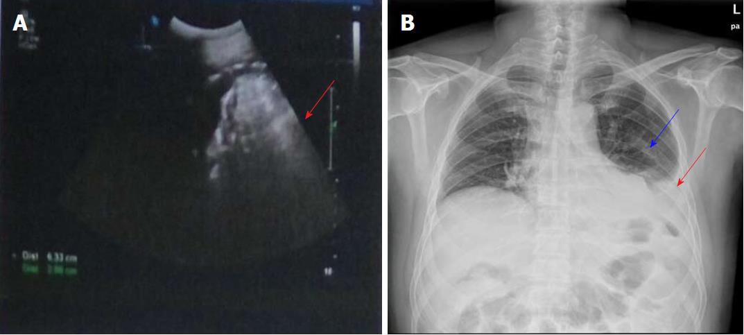
Figure 1 Abdominal B-ultrasonography findings.
A: Abdominal B-ultrasonography showed a limited non-echo area (about 6.3 cm × 2.9 cm in diameter) in the splenic fossa area, which suggested effusion in this area (arrow); B: Chest x-ray revealed a high density in the left lower chest (blue arrow), the left diaphragm angle was unclear, and a moderate volume of pleural effusion was seen in the left side of the chest (red arrow).
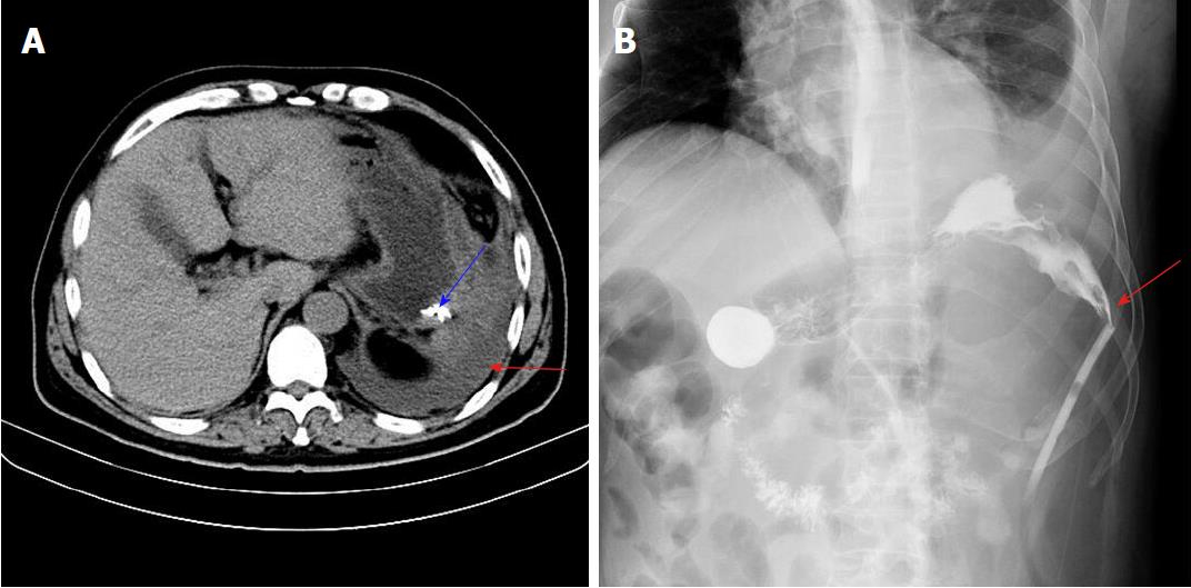
Figure 2 Upper abdominal computed tomography findings.
A: Upper abdominal computed tomography examination indicated a large amount of effusion in the left upper abdominal cavity (red arrow), and enhanced lesions and a hematocele in the drainage tube (blue arrow); B: Upper digestive tract ioversol angiography showed contrast agent overflow from the greater curvature of the gastric body to the abdominal drainage tube (arrow).
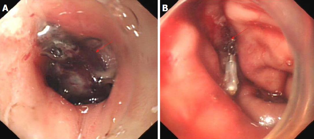
Figure 3 Gastroscopic findings.
A: Gastroscopy showed a fistula (approximately 2.0 cm × 2.5 cm in diameter) with a black blood scab on the surface of the upper side of the greater curvature of the gastric body in which the surrounding mucosa was congestive and edematous (arrow); B: The gastric body fistula was fully closed using two titanium clips guided by gastroscopy (arrow).
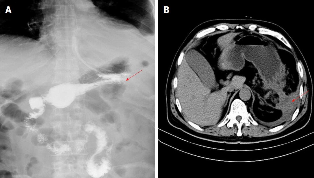
Figure 4 Upper digestive tract ioversol angiography findings.
A: Upper digestive tract ioversol angiography showed that the gastric fistula was completely closed, and no leakage of contrast medium was observed (arrow); B: Abdominal computed tomography examination showed almost no liquid retained in the abdominal cavity, and the granulation tissue was organized (arrow).
- Citation: Yu J, Zhou CJ, Wang P, Wei SJ, He JS, Tang J. Endoscopic titanium clip closure of gastric fistula after splenectomy: A case report. World J Clin Cases 2018; 6(15): 1047-1052
- URL: https://www.wjgnet.com/2307-8960/full/v6/i15/1047.htm
- DOI: https://dx.doi.org/10.12998/wjcc.v6.i15.1047


