Copyright
©The Author(s) 2018.
World J Clin Cases. Dec 6, 2018; 6(15): 1047-1052
Published online Dec 6, 2018. doi: 10.12998/wjcc.v6.i15.1047
Published online Dec 6, 2018. doi: 10.12998/wjcc.v6.i15.1047
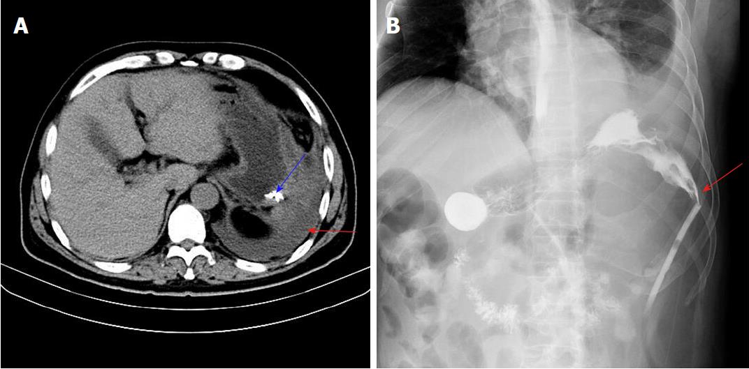
Figure 2 Upper abdominal computed tomography findings.
A: Upper abdominal computed tomography examination indicated a large amount of effusion in the left upper abdominal cavity (red arrow), and enhanced lesions and a hematocele in the drainage tube (blue arrow); B: Upper digestive tract ioversol angiography showed contrast agent overflow from the greater curvature of the gastric body to the abdominal drainage tube (arrow).
- Citation: Yu J, Zhou CJ, Wang P, Wei SJ, He JS, Tang J. Endoscopic titanium clip closure of gastric fistula after splenectomy: A case report. World J Clin Cases 2018; 6(15): 1047-1052
- URL: https://www.wjgnet.com/2307-8960/full/v6/i15/1047.htm
- DOI: https://dx.doi.org/10.12998/wjcc.v6.i15.1047


