Copyright
©2014 Baishideng Publishing Group Inc.
World J Clin Cases. May 16, 2014; 2(5): 167-169
Published online May 16, 2014. doi: 10.12998/wjcc.v2.i5.167
Published online May 16, 2014. doi: 10.12998/wjcc.v2.i5.167
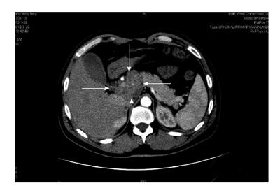
Figure 1 Computed tomography of a pancreatic tuberculosis lesion.
Computed tomography revealed a necrotic and calcified cystic lesion (white arrows) with well-defined margins in the head of the pancreas; the lesion mimicked pancreatic head carcinoma.
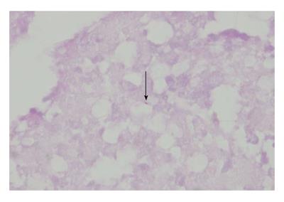
Figure 2 Histopathology of lymph node biopsies.
Ziehl-Neelsen staining of a lymph node biopsy from the pancreatic region revealed granulomatous inflammation with necrosis and acid-fast bacilli (black arrow; magnification × 400).
- Citation: Yang YJ, Li YX, Liu XQ, Yang M, Liu K. Pancreatic tuberculosis mimicking pancreatic carcinoma during anti-tuberculosis therapy: A case report. World J Clin Cases 2014; 2(5): 167-169
- URL: https://www.wjgnet.com/2307-8960/full/v2/i5/167.htm
- DOI: https://dx.doi.org/10.12998/wjcc.v2.i5.167


