Copyright
©The Author(s) 2025.
World J Clin Cases. Oct 16, 2025; 13(29): 109027
Published online Oct 16, 2025. doi: 10.12998/wjcc.v13.i29.109027
Published online Oct 16, 2025. doi: 10.12998/wjcc.v13.i29.109027
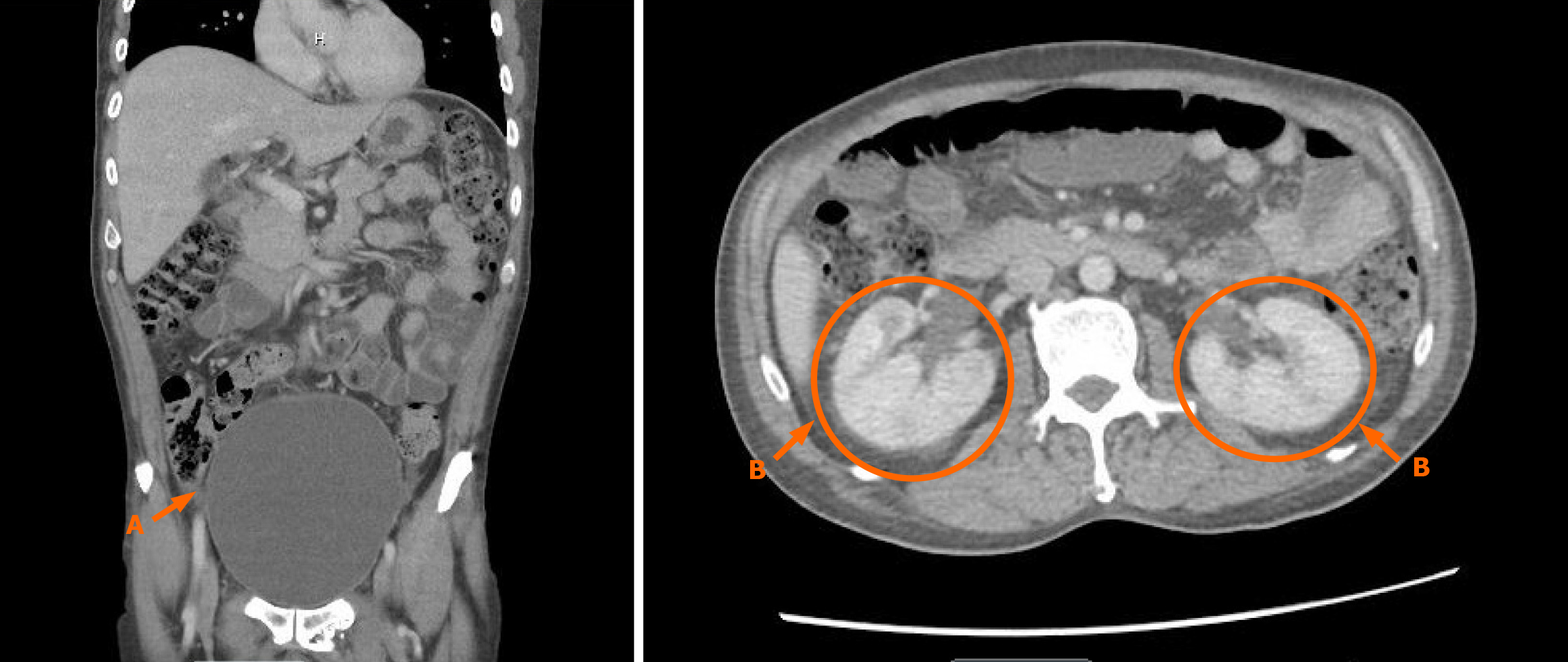
Figure 1 Computed tomography abdomen pelvis with contrast with demonstration of distended bladder (A) and perinephric stranding (B).
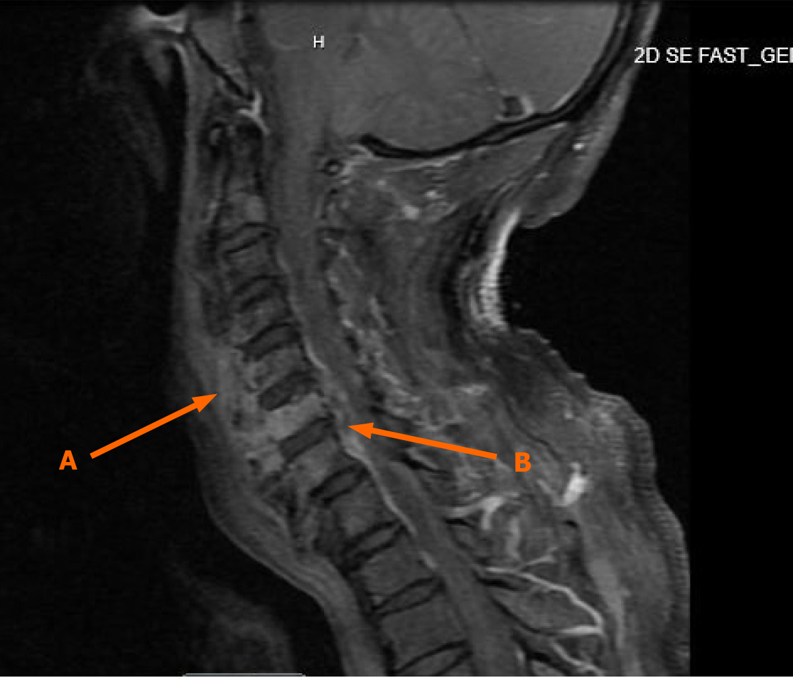
Figure 2 Magnetic resonance imaging C spine with contrast demonstrating osteomyelitis of C5-C6 (A) and C6-C7 epidural phlegmon (B).
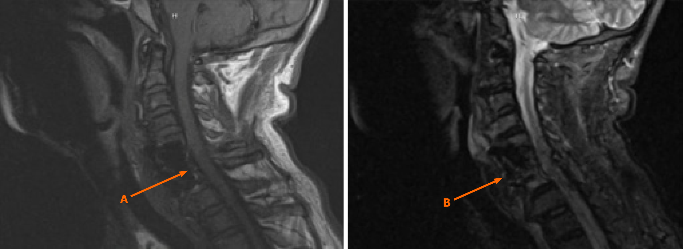
Figure 3 Magnetic resonance imaging C spine with and without contrast showed no evidence of discitis, osteomyelitis or epidural abscess (A and B).
- Citation: AlSabea N, Kanor U, Garcia AS, Shah A, Sun A. Spinal epidural abscess of uncommon presentation following urinary tract infection: A case report. World J Clin Cases 2025; 13(29): 109027
- URL: https://www.wjgnet.com/2307-8960/full/v13/i29/109027.htm
- DOI: https://dx.doi.org/10.12998/wjcc.v13.i29.109027


