Copyright
©The Author(s) 2025.
World J Clin Cases. Oct 16, 2025; 13(29): 108608
Published online Oct 16, 2025. doi: 10.12998/wjcc.v13.i29.108608
Published online Oct 16, 2025. doi: 10.12998/wjcc.v13.i29.108608
Figure 1 Computed tomography.
A: Computed tomography (CT) angiography after first transplant procedure showed a lack of opacification in the hepatic arteries; B: CT scan after first transplant showed localized ischemic zones in segment VI; C: Subacute and acute peripheral wedge infarcts of segments VI and VII of the liver graft on follow-up CT scan.
Figure 2 Surgical procedure details.
A: The first transplanted graft with visible areas of ischemia due to hepatic artery thrombosis; B: The anastomosis of the donor aorta and celiac trunk as a patch on the recipient’s supraceliac aorta; C: The placement of the tissue expander between the liver and right abdominal wall.
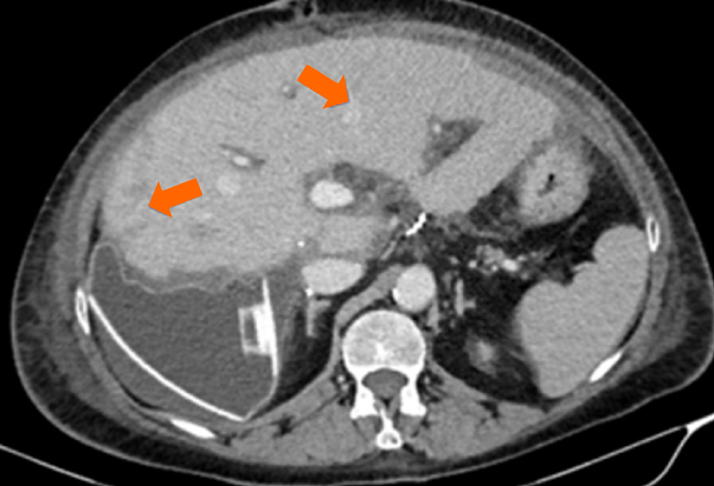
Figure 3 Follow-up computed tomography scan.
Ischemic areas in segments VII and IVa of the liver graft were not clinically relevant.
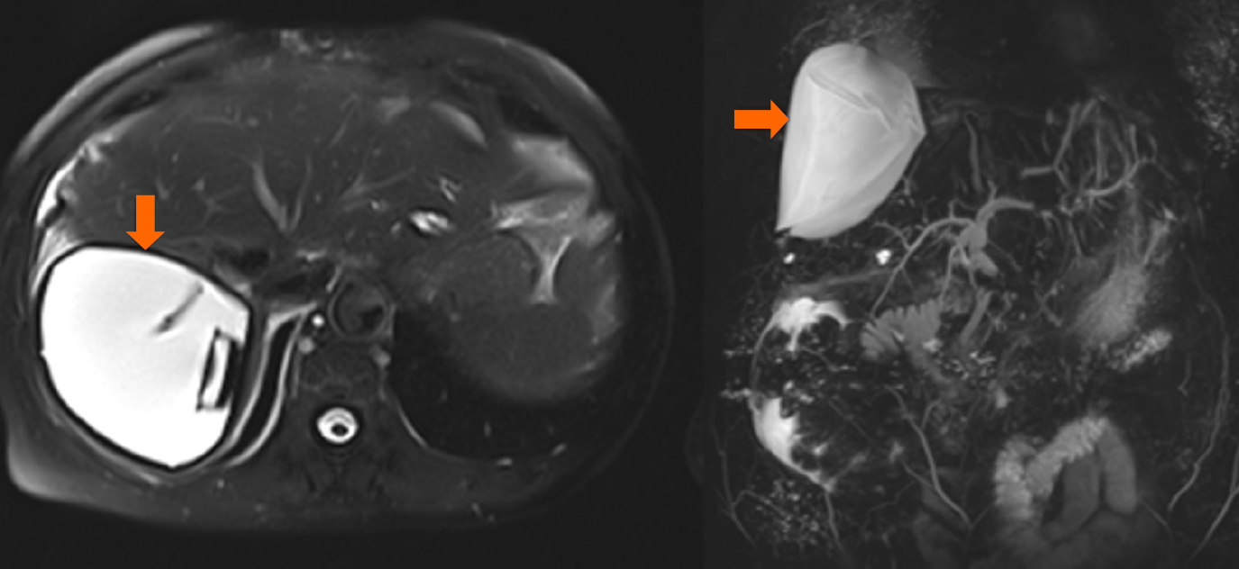
Figure 4 Follow-up magnetic resonance imaging.
Optimal position of the tissue expander with patent vessels and no new ischemic lesions.
- Citation: Petrovic I, Brekalo K, Romic I, Silovski H, Sesa V, Galunic Cicak R, Bulic K, Koltay OM, Strajher IM, Mrzljak A. Tissue expanders – an innovative approach in the management of hepatic venous outflow obstruction after liver transplantation: A case report. World J Clin Cases 2025; 13(29): 108608
- URL: https://www.wjgnet.com/2307-8960/full/v13/i29/108608.htm
- DOI: https://dx.doi.org/10.12998/wjcc.v13.i29.108608


