Copyright
©The Author(s) 2025.
World J Clin Cases. Aug 26, 2025; 13(24): 107825
Published online Aug 26, 2025. doi: 10.12998/wjcc.v13.i24.107825
Published online Aug 26, 2025. doi: 10.12998/wjcc.v13.i24.107825
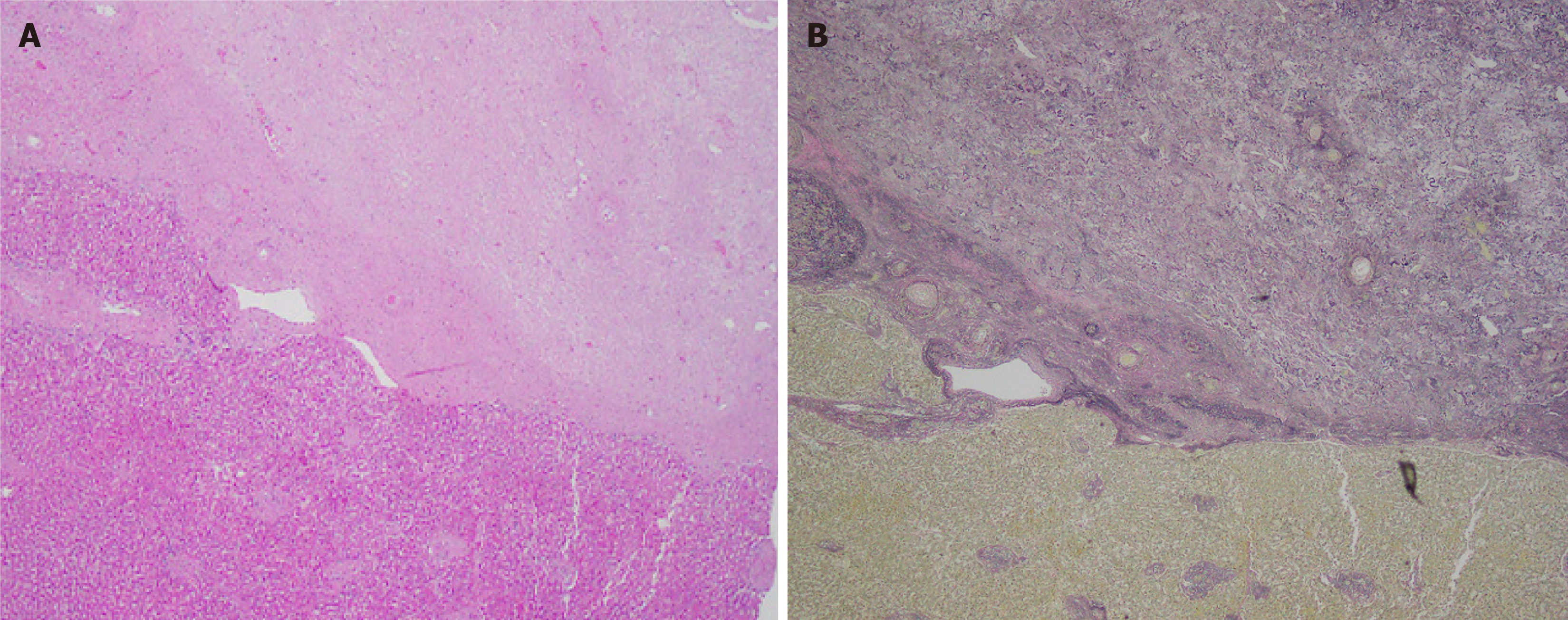
Figure 1 Segmental atrophy of liver: Identified in a 79-year-old male who presented with abdominal bloating.
A: An abrupt interface exists between the lesion and the background liver. The lesion, in its elastotic stage, is composed almost entirely of elastic-rich matrix (hematoxylin-eosin, ×40); B: Elastin staining highlights the elastic fibers (Elastin, ×40).
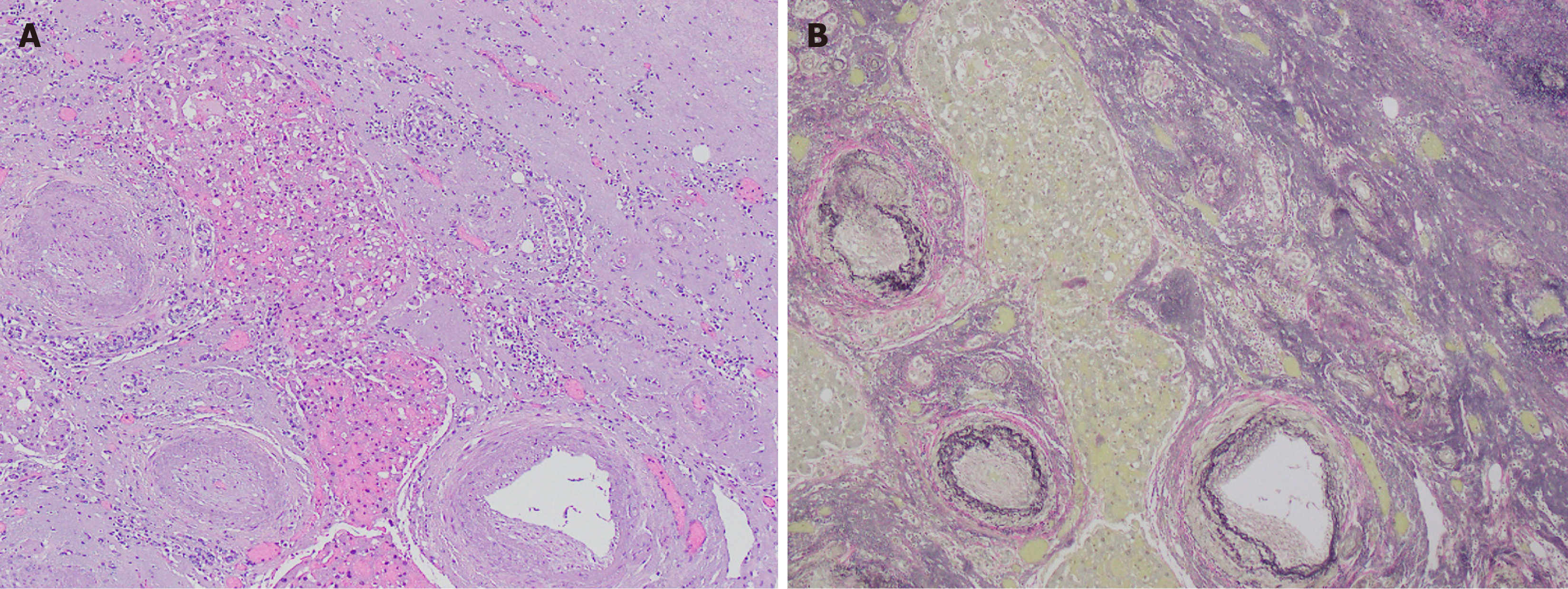
Figure 2 Segmental atrophy of liver: An incidental finding in the autopsy of an 87-year-old female died of cardiopulmonary arrest.
A: The lesion consists of an elastic-rich matrix with entrapped islands of hepatocytes. The abnormally thick-walled vessels with fibrosis are striking (hematoxylin-eosin, ×100); B: Elastin staining highlights the extensive elastosis as well as blood vessels (Elastin, ×100).
- Citation: Younus A, Liu Y, Connor EE, Wu ZY, Lee H, Fu ZY. Segmental atrophy of the liver: Review of a rare pseudotumor. World J Clin Cases 2025; 13(24): 107825
- URL: https://www.wjgnet.com/2307-8960/full/v13/i24/107825.htm
- DOI: https://dx.doi.org/10.12998/wjcc.v13.i24.107825


