Copyright
©The Author(s) 2025.
World J Clin Cases. Aug 16, 2025; 13(23): 106329
Published online Aug 16, 2025. doi: 10.12998/wjcc.v13.i23.106329
Published online Aug 16, 2025. doi: 10.12998/wjcc.v13.i23.106329
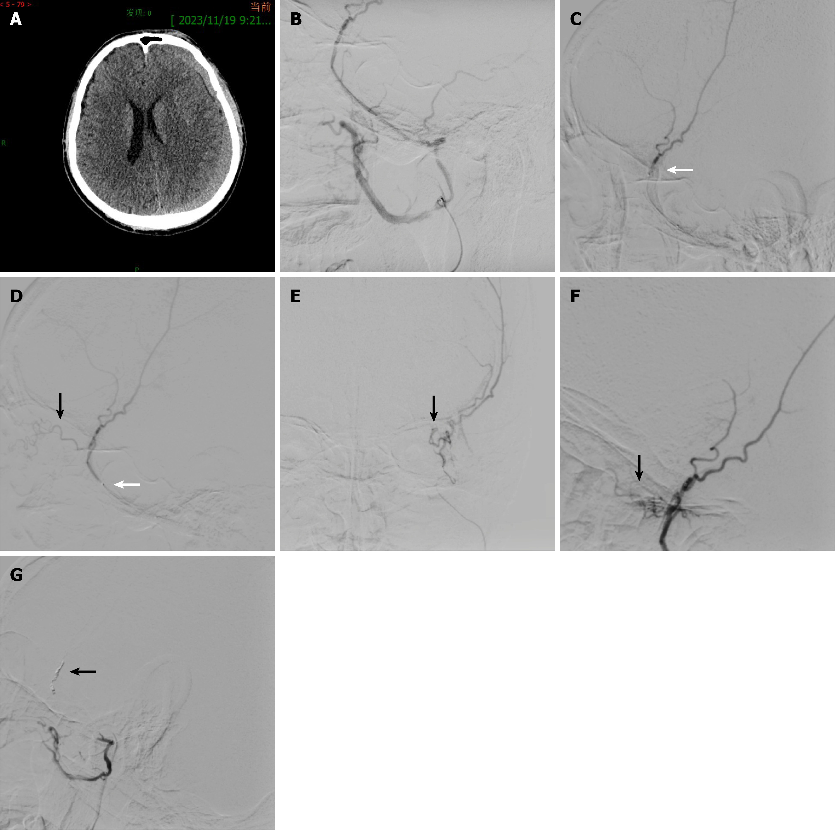
Figure 1 Computed tomography and angiography.
A: Brain computed tomography scan shows left chronic subdural hematoma; B: Middle meningeal artery (MMA) angiography revealed absence of detectable anastomotic artery along the anterior trunk; C: The tip of the microcatheter (white arrow) was placed distal to the main trunk of the anterior branch of the MMA for polyvinyl alcohol (PVA) injection; D: Lateral imaging of the microcatheter tip (white arrow) retracted due to intraoperative patient movement, revealing the anastomotic artery (black arrow) upon PVA injection; E: Anteroposterior view of PVA flowing through the long, tortuous sphenoidal artery (black arrow) to the lacrimal artery, which supplies the outer superior quadrant of the orbit; F: Reflux of PVA into the anastomotic artery (black arrow) during reinjection; G: Post-embolization contrast showing complete occlusion of the anterior and posterior branches of the MMA, with coils visible in the anterior branch (black arrow).
- Citation: Zhao F, Su CH, Hu SX, Feng L. Diplopia after middle meningeal artery embolization for chronic subdural hematoma: A case report. World J Clin Cases 2025; 13(23): 106329
- URL: https://www.wjgnet.com/2307-8960/full/v13/i23/106329.htm
- DOI: https://dx.doi.org/10.12998/wjcc.v13.i23.106329


