Copyright
©The Author(s) 2025.
World J Clin Cases. Aug 16, 2025; 13(23): 106321
Published online Aug 16, 2025. doi: 10.12998/wjcc.v13.i23.106321
Published online Aug 16, 2025. doi: 10.12998/wjcc.v13.i23.106321
Figure 1 Venous blood sample from the patient.
The blood was visually lactescent.
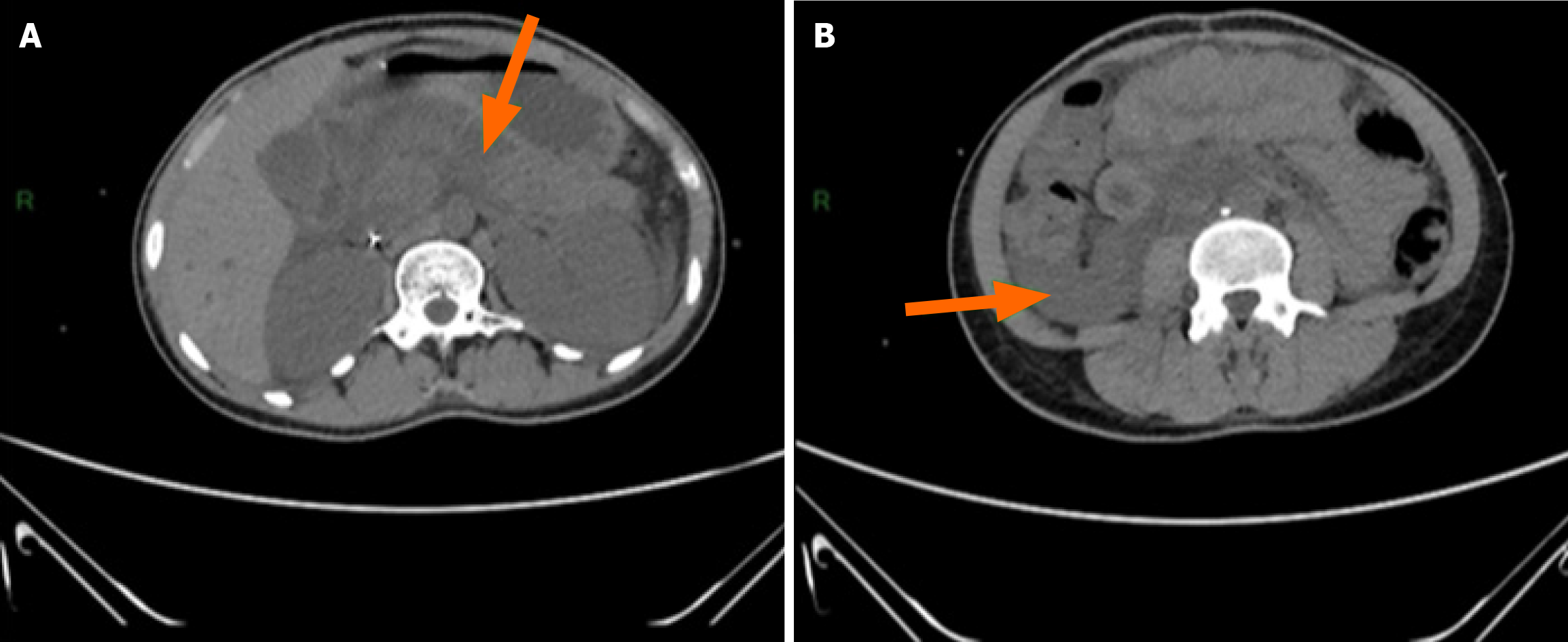
Figure 2 Computed tomography imaging findings.
A: Pancreatic flow (arrow); B: Thickening of the pancreas with pancreatic flow (arrow).
- Citation: Kouame KI, Mobio PMN, Bouh JK, Konan JK, Coulibaly TK, Toure CW, Diebi LAA, Kouakou JNH, Koffi BE, Yapo PY. Convergence of diabetic ketoacidosis, acute pancreatitis, and malaria: A case report. World J Clin Cases 2025; 13(23): 106321
- URL: https://www.wjgnet.com/2307-8960/full/v13/i23/106321.htm
- DOI: https://dx.doi.org/10.12998/wjcc.v13.i23.106321


