Copyright
©The Author(s) 2025.
World J Clin Cases. Aug 16, 2025; 13(23): 105671
Published online Aug 16, 2025. doi: 10.12998/wjcc.v13.i23.105671
Published online Aug 16, 2025. doi: 10.12998/wjcc.v13.i23.105671
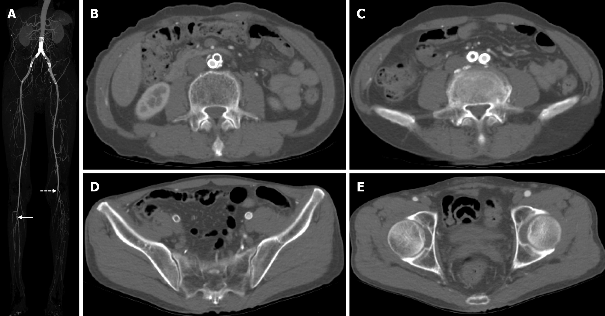
Figure 1 Preoperative imaging.
A: Preoperative maximal intensity projection computed tomography image demonstrated the stent placement from the abdominal aorta to the external iliac artery just above the circumflex iliac artery. And there was minimal grade calcification in the right tibioperoneal trunk (arrow) and complete occlusion of the left popliteal artery (dotted arrow); B-D: Preoperative axial computed tomography images showed the thrombotic occlusion of the stent at the level of the abdominal aorta, right common iliac artery, and right external iliac artery; E: Continued image showed the patent right common femoral artery.
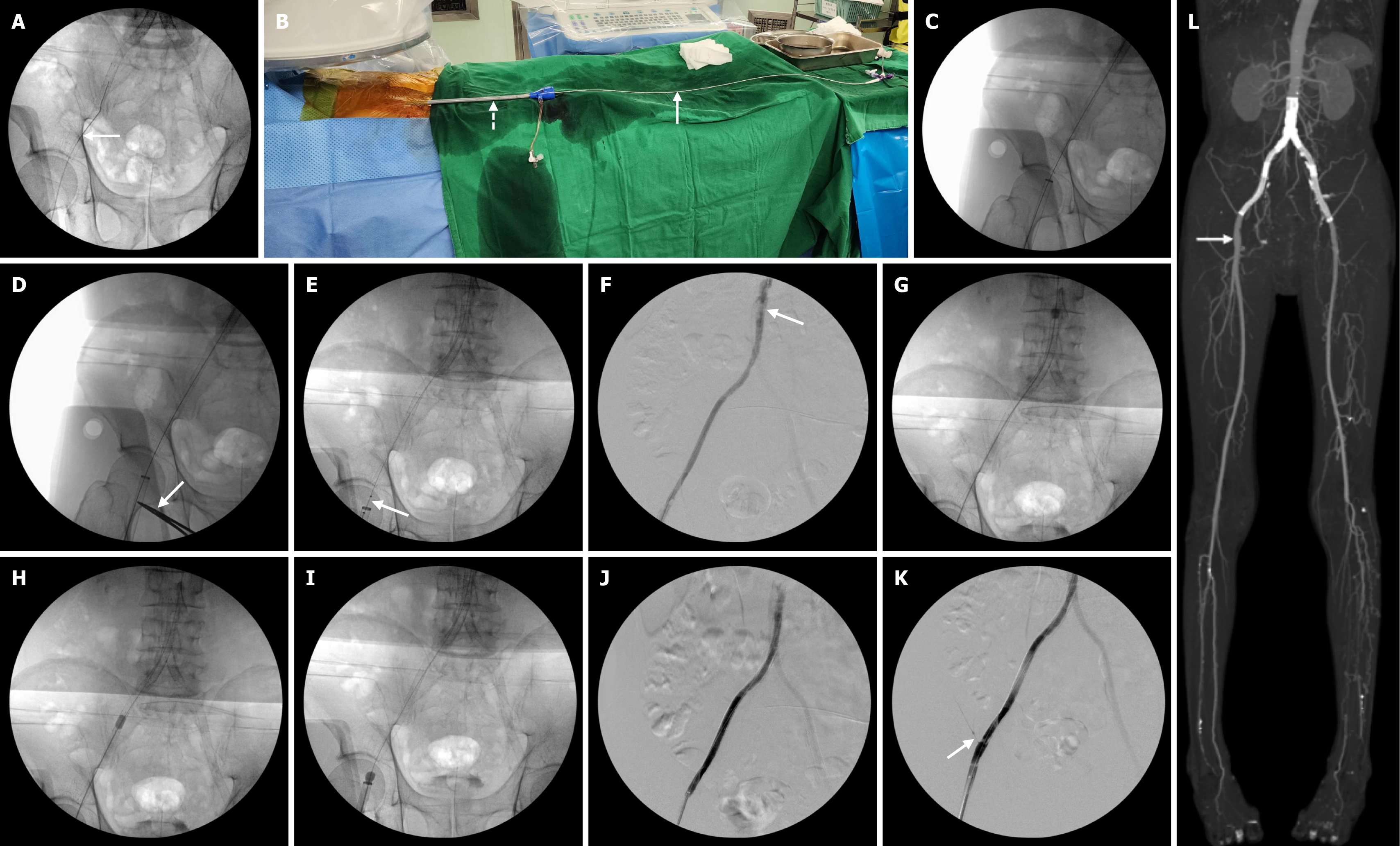
Figure 2 Procedure details of the percutaneous treatment for the thrombotic occlusion of iliac artery stent.
A: An 035In straight-tip guidewire was inserted through the right common femoral artery (arrow); B: After a successful preclose technique using two 6F Proglides, a 22 French Sentrant large bore sheath was inserted into the right common femoral artery to prevent distal embolization during the procedure (dotted arrow). Then, an Angiojet thrombectomy catheter was introduced through this sheath (arrow); C: Fluoroscopic image showed a guidewire passage and placement of a large bore sheath; D: Marking the access site using the placement of a mosquito clamp could be helpful to inadvertent pullout of a large bore sheath (arrow); E: Aspirational thrombectomy was performed using an Angiojet thrombectomy catheter with retrograde fashion (arrow); F: After Angiojet thrombectomy, the angiogram demonstrated a nearly complete thrombectomy and residual organized thrombus at the proximal stent portion (arrow); G-I: Conventional mechanical thrombectomy was done using a 5.5 Fr over-the-wire Fogarty catheter from the proximal portion of the stent to the tip of the sheath. Eventually, the organized thrombus was collected in the large bore sheath; J and K: The completion angiogram showed the complete removal of the thrombus and patent stent including the circumflex iliac artery (arrow); L: Postoperative maximal intensity projection computed tomography image showed no evidence of distal embolization.
- Citation: Cho S, Joh JH. Retrograde approach of Angiojet catheter for the acute occlusion of aortoiliac artery stent: A case report. World J Clin Cases 2025; 13(23): 105671
- URL: https://www.wjgnet.com/2307-8960/full/v13/i23/105671.htm
- DOI: https://dx.doi.org/10.12998/wjcc.v13.i23.105671


