Copyright
©The Author(s) 2025.
World J Clin Cases. Aug 16, 2025; 13(23): 104807
Published online Aug 16, 2025. doi: 10.12998/wjcc.v13.i23.104807
Published online Aug 16, 2025. doi: 10.12998/wjcc.v13.i23.104807
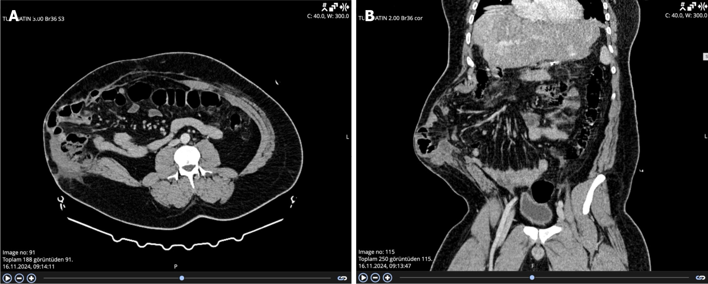
Figure 1 Computed tomography images.
A: Computed tomography (CT) images of abdominal hernia in axial section; B: CT images of abdominal hernia in coronal section.
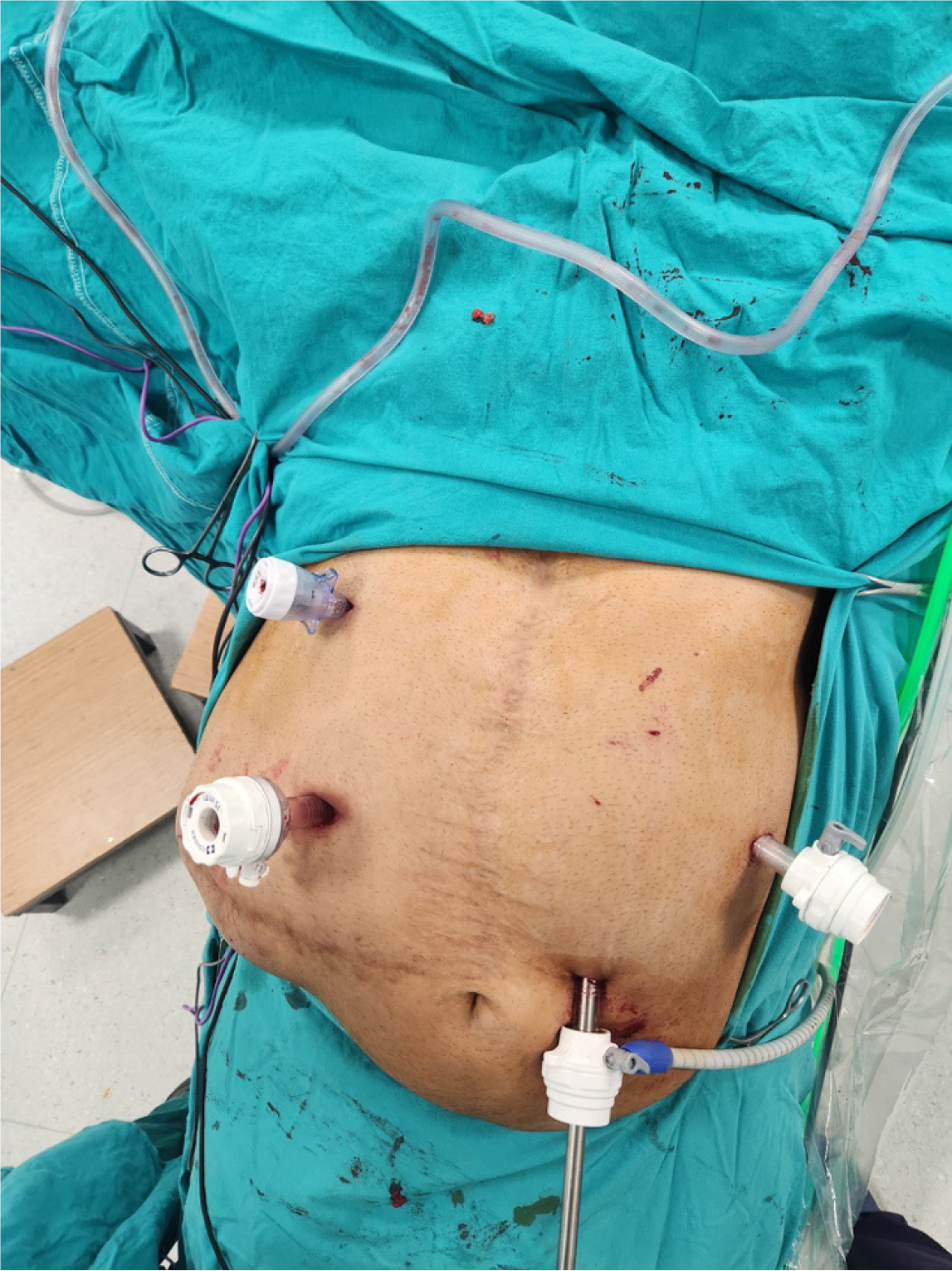
Figure 2 Locations of the trocars.
(C) Camera port (10 mm) inserted 2 cm left to the umbilicus; (W1) Working port 1 (15 mm) inserted at the midclavicular line in the right upper quadrant; (W2) Working port 2 (12 mm) inserted at the anterior axillary line on the left; (A) Assistance port (5 mm) inserted at the intersection of the midclavicular line and the subcostal margin.
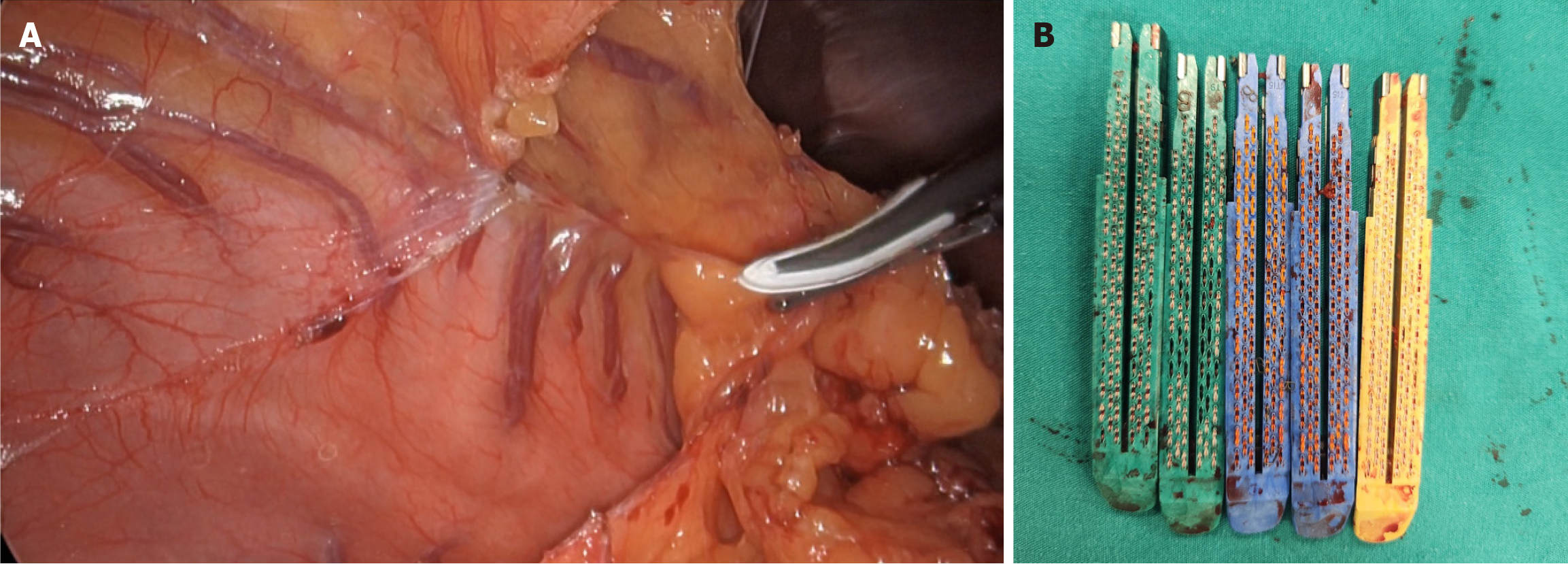
Figure 3 Laparoscopic view and total number and sizes of cartridges used during sleeve gastrectomy.
A: Dissection of the lesser omentum by division of the gastrocolic ligament; B: The number and size of endoscopic cartridges used during laparoscopic sleeve gastrectomy.
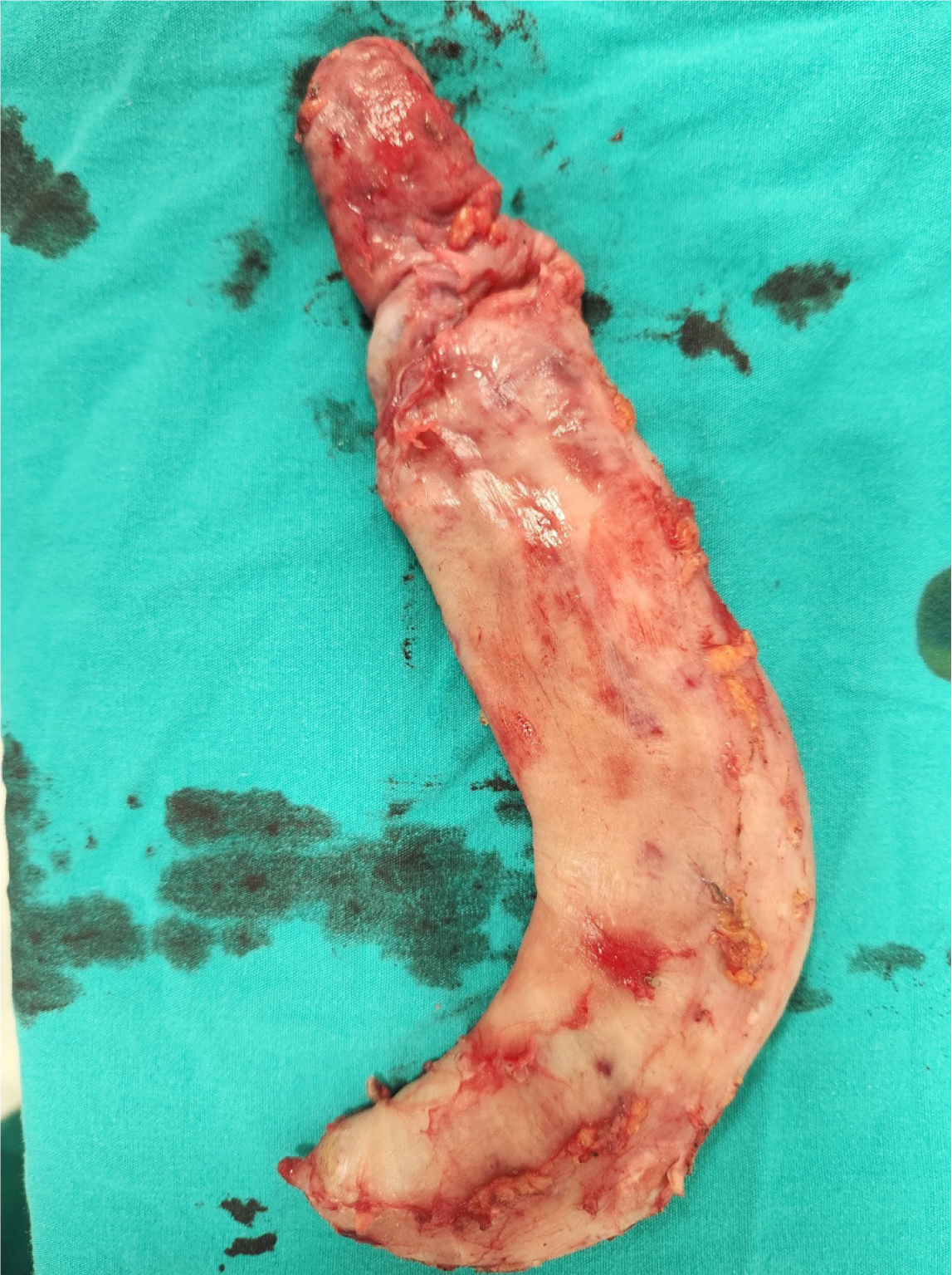
Figure 4
The surgical specimen.
- Citation: Cicek E, Karatepe YK, Kantarcı TR, Sahin TT. Demanding sleeve gastrectomy procedure in a patient with severe intraabdominal adhesions: A case report and review of the literature. World J Clin Cases 2025; 13(23): 104807
- URL: https://www.wjgnet.com/2307-8960/full/v13/i23/104807.htm
- DOI: https://dx.doi.org/10.12998/wjcc.v13.i23.104807


