Copyright
©The Author(s) 2025.
World J Clin Cases. Aug 6, 2025; 13(22): 104352
Published online Aug 6, 2025. doi: 10.12998/wjcc.v13.i22.104352
Published online Aug 6, 2025. doi: 10.12998/wjcc.v13.i22.104352
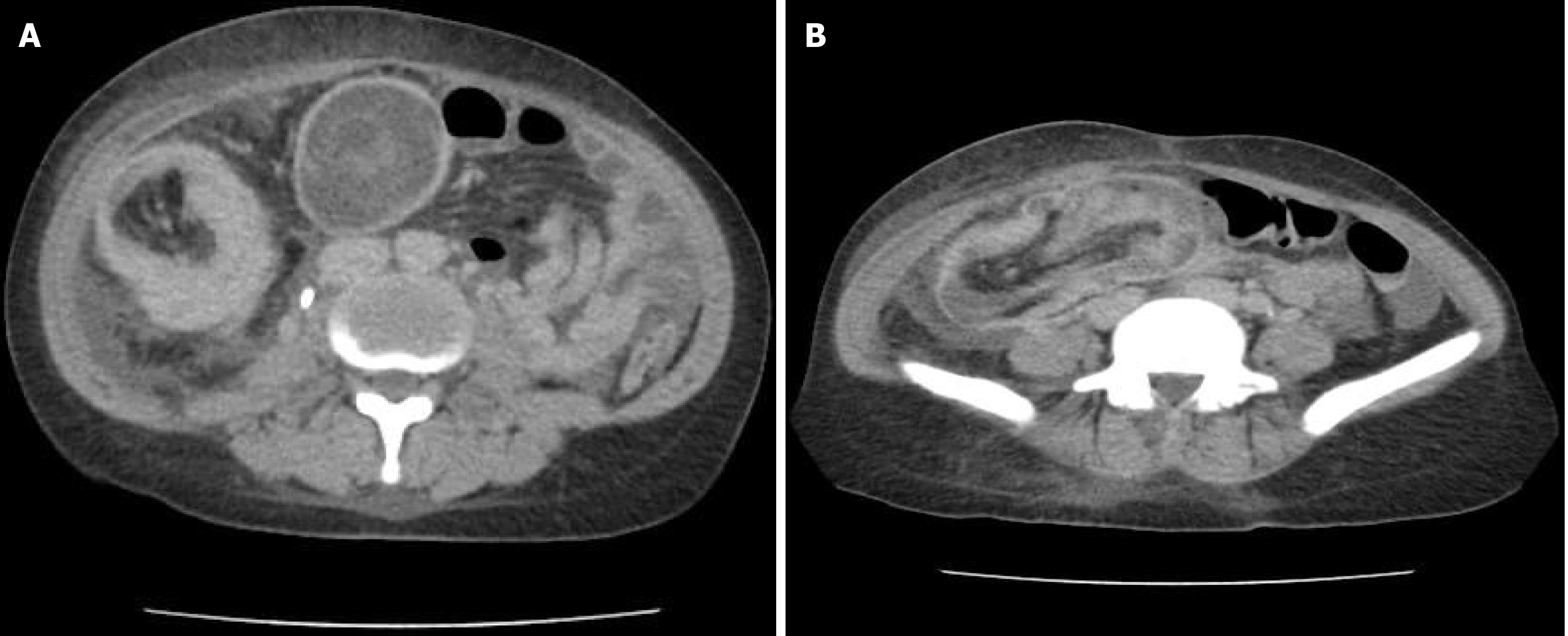
Figure 1 Computed tomography abdominal scan.
A: A mass in the cecum and ascending colon; B: Ileal invagination into the cecum.
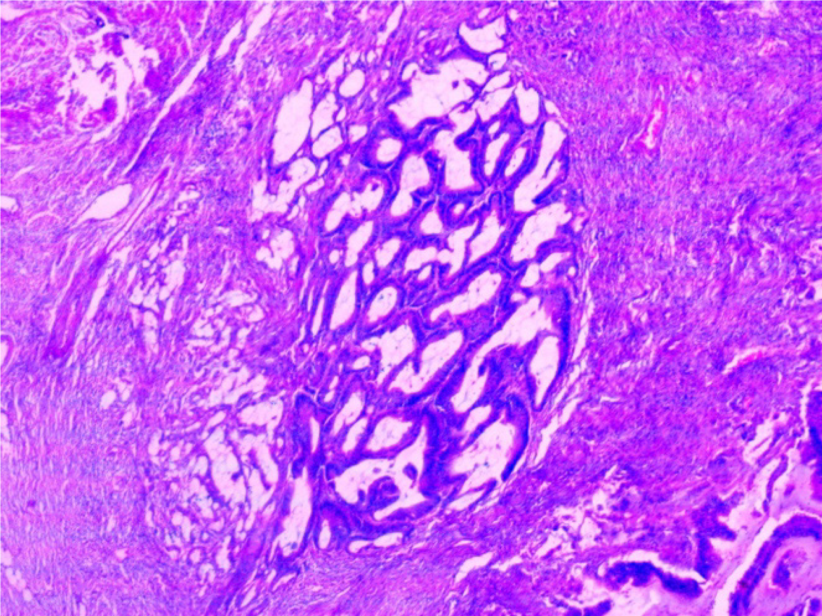
Figure 2
Histopathological micrograph showing polygonal cells with variable mucin and glandular debris invading the mucosa.
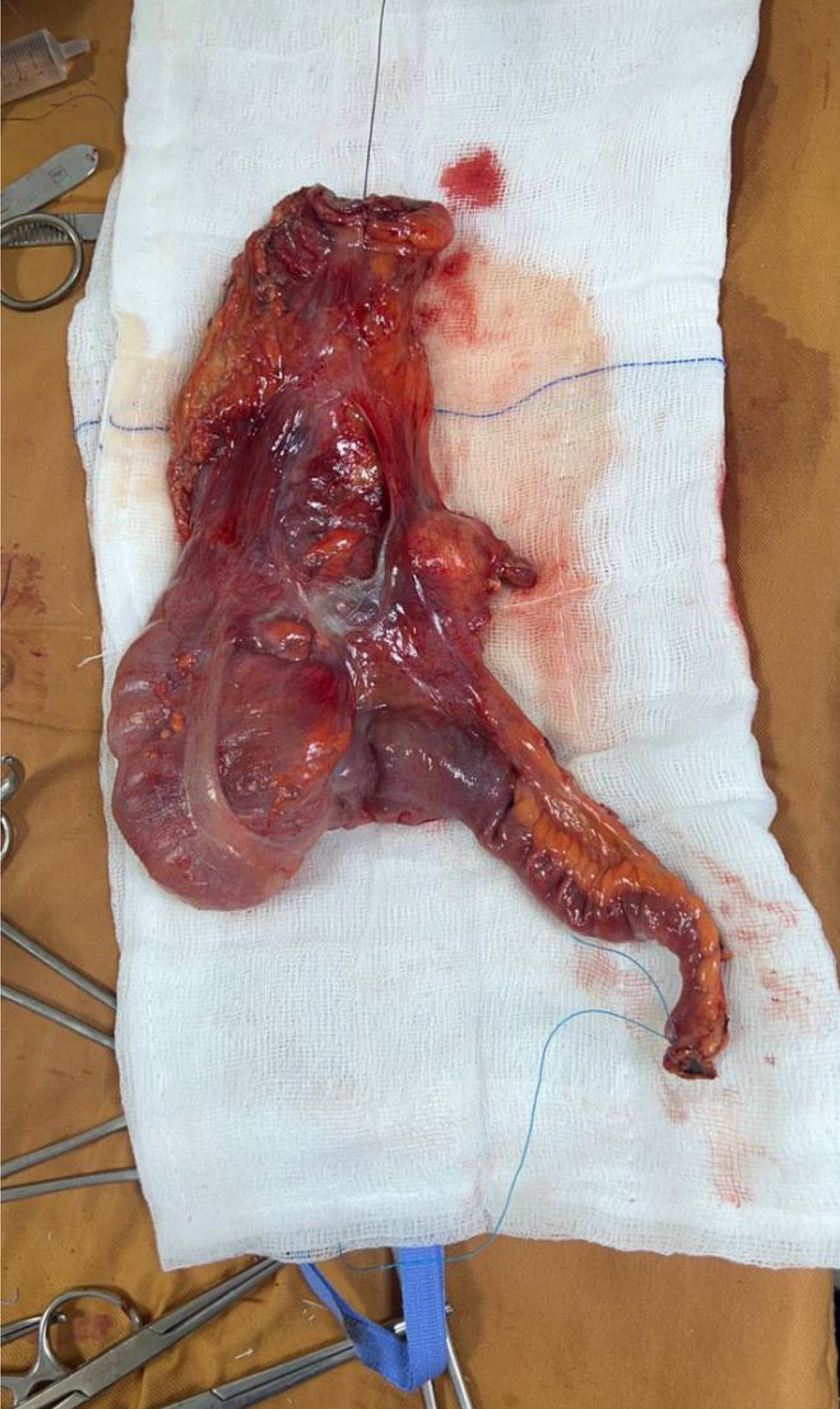
Figure 3
Resected portion of the ileum, cecum, and ascending colon.
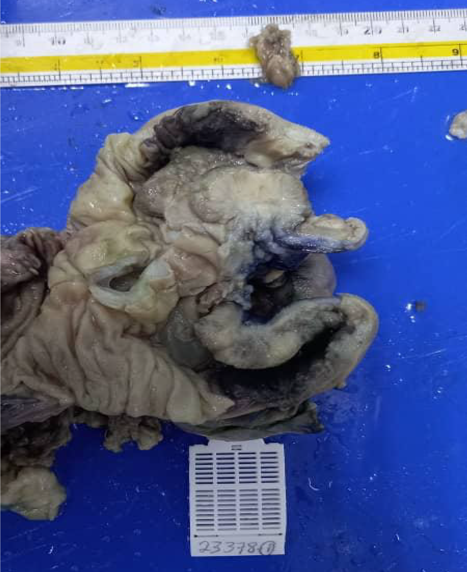
Figure 4
Gross pathological description showing a large polypoid mass in the cecum and ascending colon.
- Citation: Abdishakur AE, Ahmed MAA. Adult ileo cecal intussusception as a manifestation of colon carcinoma: A case report. World J Clin Cases 2025; 13(22): 104352
- URL: https://www.wjgnet.com/2307-8960/full/v13/i22/104352.htm
- DOI: https://dx.doi.org/10.12998/wjcc.v13.i22.104352


