Copyright
©The Author(s) 2024.
World J Clin Cases. Apr 6, 2024; 12(10): 1817-1823
Published online Apr 6, 2024. doi: 10.12998/wjcc.v12.i10.1817
Published online Apr 6, 2024. doi: 10.12998/wjcc.v12.i10.1817
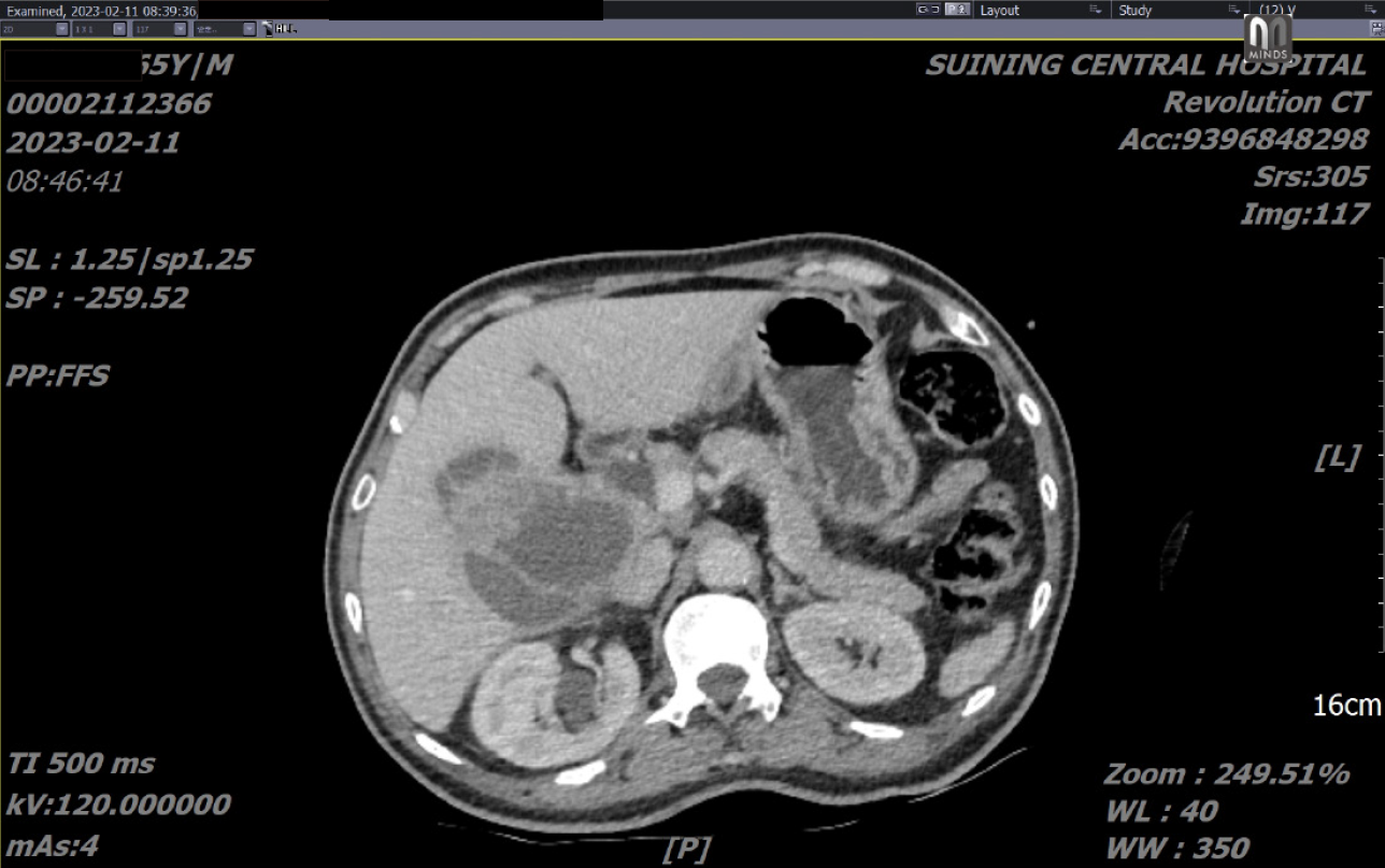
Figure 1 Enhanced computed tomography scan revealed a polycystic mass of 10.
1 cm × 7.5 cm in size in the right liver and gallbladder fossa with a thick and enhanced cyst wall. Gallbladder was not obvious. No evidence of gallstones were seen.
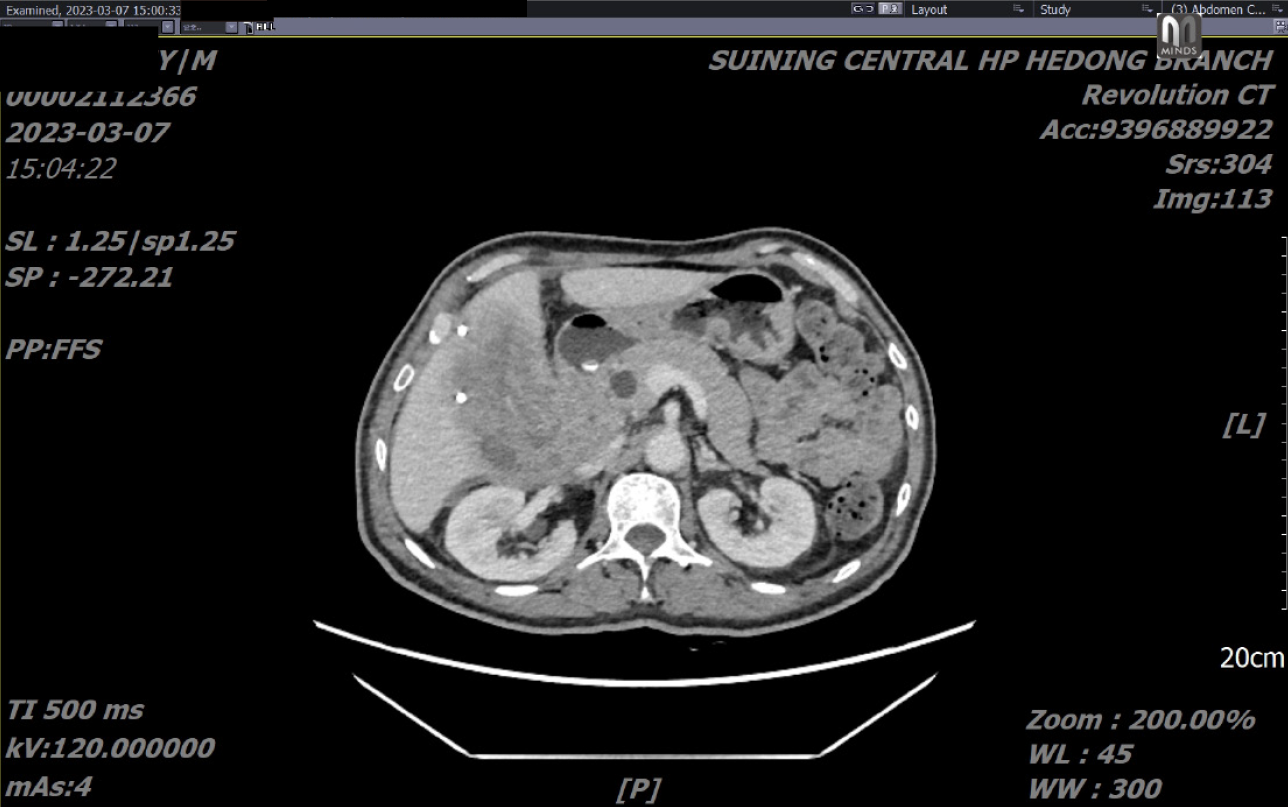
Figure 2 A month later, computed tomography reexamination revealed that the liver mass had not shrunk significantly and had become a fully solid mass.
The common bile duct is not dilated by the pressure.
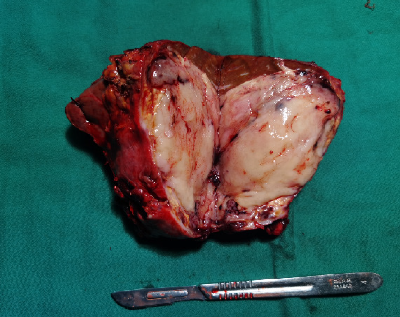
Figure 3 The resected specimen was a nodular mass with a size of about 9 cm × 8 cm × 6.
5 cm. The section is grayish white, solid tissue, soft texture, and adhesion to the surrounding tissue. The gallbladder was invaded into the liver. The gallbladder contained new fish-like tissue. Necrosis and bleeding were seen in the lesion.
Figure 4 Pathological features of carcinosarcoma of gallbladder.
A: High power view of the gallbladder wall show intramucosal adenocarcinoma [hematoxylin and eosin (H&E) stain, 40 ×]; B: High power view show high-grade spindle cell sarcoma (H&E, 40 ×); C: Cytokeratin (CK) (HIC, 40 ×) staining show strong membranous positivity in the intramucosal adenocarcinoma; D: CK-19 (HIC, 10 ×) staining show strong membranous positivity in the intramucosal adenocarcimoma; E: Transducin-like enhancer protein 1 (HIC, 40 ×) staining show strong sarcomatous component positivity in the gallbladder neoplasm; F: Special AT-rich sequence-binding protein 2 (HIC, 40 ×) staining show osteoblastic component positivity in the gallbladder neoplasm.
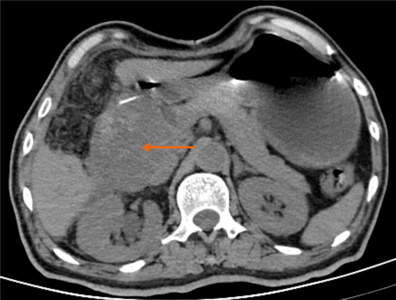
Figure 5 One month after surgery, the patient had recurrent episodes of nausea and vomiting.
Vomiting more stomach contents. Abdominal computed tomography (CT) scan (the patient refused to undergo enhanced CT) revealed a 9 cm × 8 cm × 8 cm mass in the anterior duodenum (indicated by the red arrow), leading to duodenal obstruction. Local recurrence of the tumor is suspected.
- Citation: Dai Y, Meng M, Luo QZ, Liu YJ, Xiao F, Wang CH. Gallbladder carcinosarcoma with a poor prognosis: A case report. World J Clin Cases 2024; 12(10): 1817-1823
- URL: https://www.wjgnet.com/2307-8960/full/v12/i10/1817.htm
- DOI: https://dx.doi.org/10.12998/wjcc.v12.i10.1817


