Copyright
©The Author(s) 2024.
World J Clin Cases. Apr 6, 2024; 12(10): 1799-1803
Published online Apr 6, 2024. doi: 10.12998/wjcc.v12.i10.1799
Published online Apr 6, 2024. doi: 10.12998/wjcc.v12.i10.1799
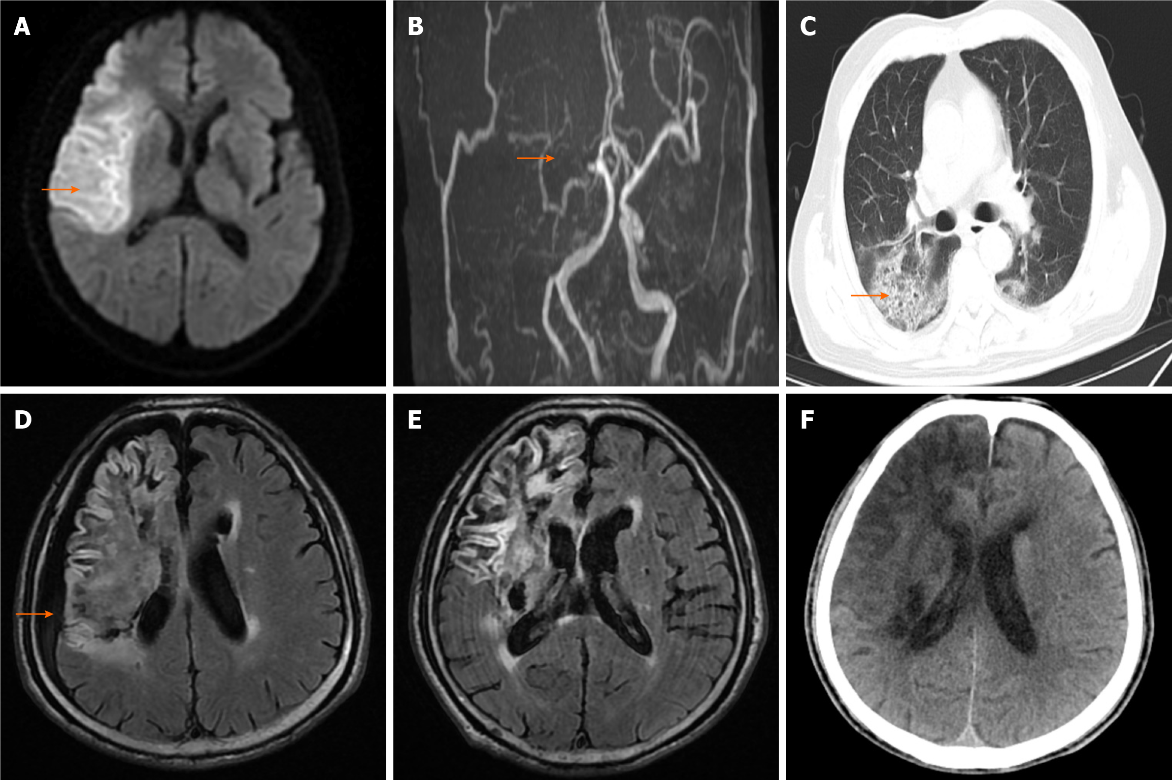
Figure 1 Cranial magnetic resonance imaging.
A: Cranial magnetic resonance imaging (MRI) revealed distinct, patchy, nodular long T2 signal shadows in the right frontal, temporal, and parietal lobes; B: Head and neck MRI displayed non-visualization of the right internal carotid and middle cerebral arteries, particularly noting superficial shadows in the right internal carotid siphon, the middle cerebral artery M1 segment, and the right anterior cerebral artery A1 segment (indicated by arrow); C: Chest computed tomography showed scattered patchy shadows in the lungs; D: Cranial MRI displayed new subdural effusion in the right frontal, temporal, and parietal regions; E: Cranial MRI displayed marked absorption of subdural effusion in the right frontal, temporal, and parietal regions at 6 d after its appearance; F: Head computed tomography displayed the scar of cerebral infarction with hemorrhagic transformation in the right frontal, temporal, and parietal lobes at 3 months after discharge.
- Citation: Xue ZY, Xiao ZL, Cheng M, Xiang T, Wu XL, Ai QL, Wu YL, Yang T. Subdural effusion associated with COVID-19 encephalopathy: A case report. World J Clin Cases 2024; 12(10): 1799-1803
- URL: https://www.wjgnet.com/2307-8960/full/v12/i10/1799.htm
- DOI: https://dx.doi.org/10.12998/wjcc.v12.i10.1799


