Copyright
©The Author(s) 2023.
World J Clin Cases. Mar 26, 2023; 11(9): 2009-2014
Published online Mar 26, 2023. doi: 10.12998/wjcc.v11.i9.2009
Published online Mar 26, 2023. doi: 10.12998/wjcc.v11.i9.2009
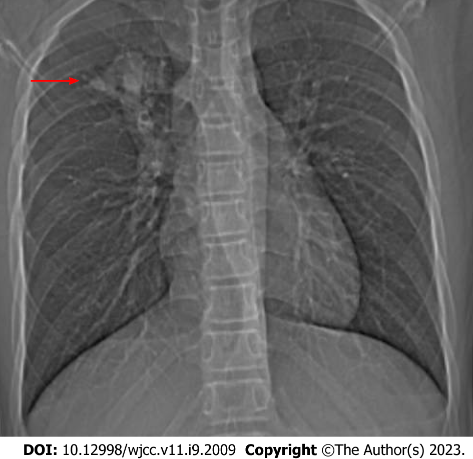
Figure 1 Chest x-ray.
A mass shadow in the right upper lung (red arrow).
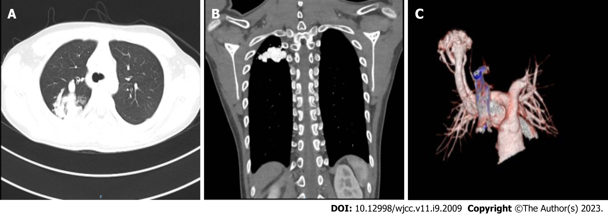
Figure 2 Cardiovascular computed tomography angiography.
A and B: Representative images showing the abnormal vascular nest in the right upper lung; C: Expansion of the right upper pulmonary artery with thickened and twisted branching vessels to form an abnormal vascular nest with direct reflux into the right upper pulmonary posterior vein. The artery finally merged into the right upper pulmonary vein.
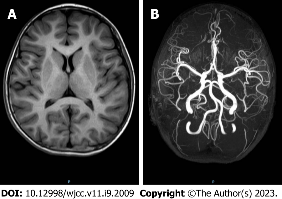
Figure 3 Brain magnetic resonance imaging and cerebral magnetic resonance angiography.
A: Magnetic resonance imaging showed no obvious abnormalities; B: Magnetic resonance angiography showed no obvious abnormalities.
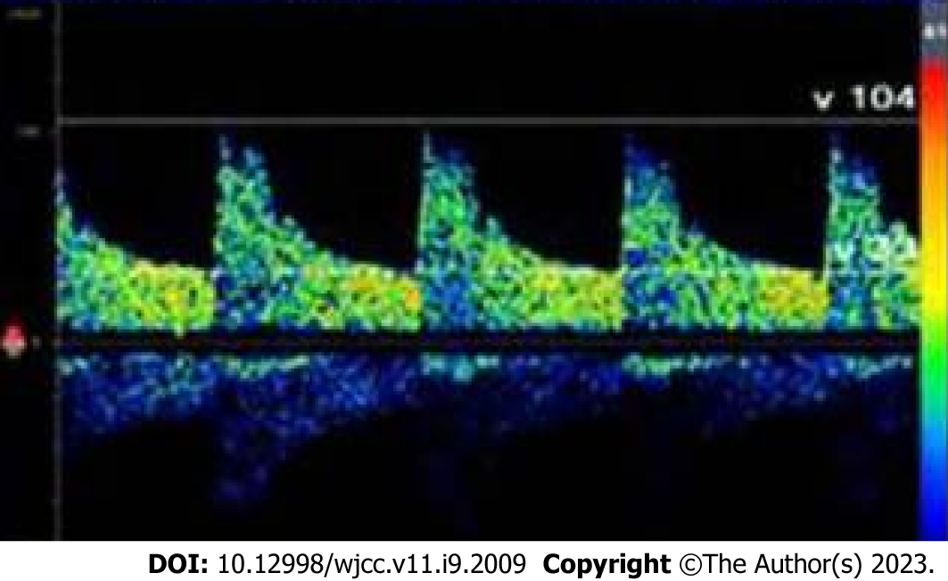
Figure 4 Contrast-enhanced transcranial Doppler ultrasound.
Significant embolus signals appeared in the middle cerebral artery within 10 s after Valsalva maneuver.
Figure 5 Angiography and embolization treatment.
A and B: Representative angiography images showing a pulmonary arteriovenous fistula (PAVF) originating from the right upper pulmonary branch artery; C and D: Representative images showing the PAVF was no longer detected after embolization with a vascular plug.
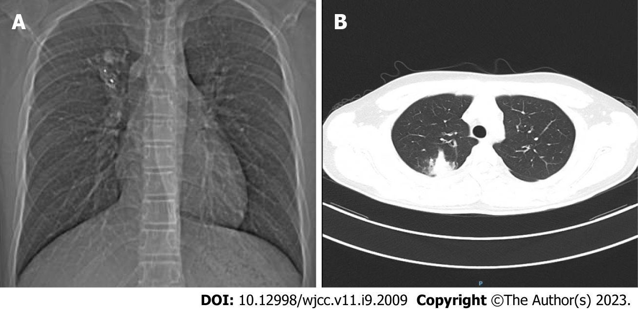
Figure 6 Postoperative follow-up imaging.
A: Chest x-ray showed that the position of the vascular plug was stable; B: Computed tomography showed that the pulmonary arteriovenous fistula markedly shrank after embolization therapy.
- Citation: Zheng J, Wu QY, Zeng X, Zhang DF. Transient ischemic attack induced by pulmonary arteriovenous fistula in a child: A case report. World J Clin Cases 2023; 11(9): 2009-2014
- URL: https://www.wjgnet.com/2307-8960/full/v11/i9/2009.htm
- DOI: https://dx.doi.org/10.12998/wjcc.v11.i9.2009


