Copyright
©The Author(s) 2023.
World J Clin Cases. Mar 26, 2023; 11(9): 1974-1984
Published online Mar 26, 2023. doi: 10.12998/wjcc.v11.i9.1974
Published online Mar 26, 2023. doi: 10.12998/wjcc.v11.i9.1974
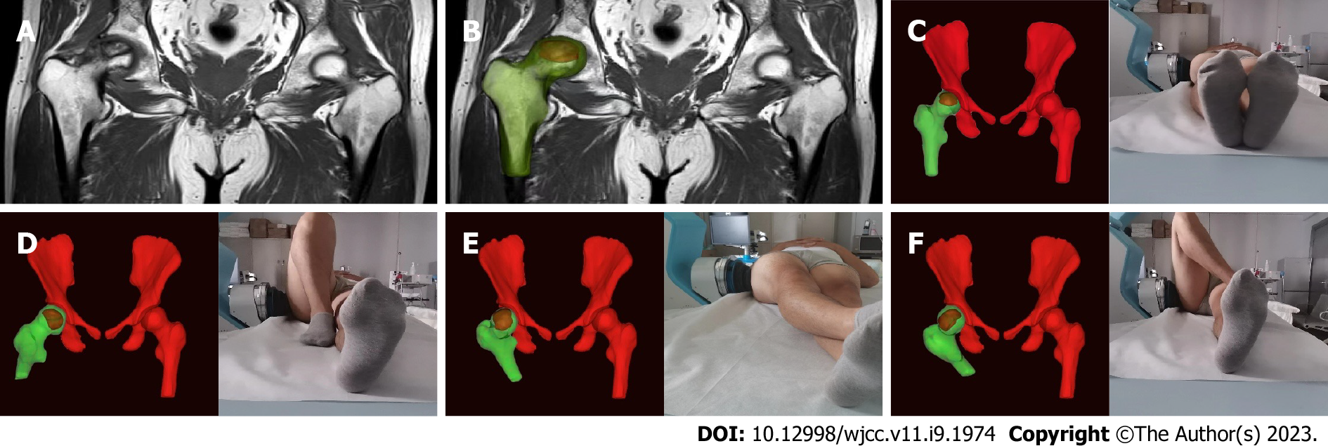
Figure 1 Personalized extracorporeal shock wave therapy based on magnetic resonance imaging three-dimensional reconstruction.
A: Magnetic resonance imaging (MRI) image; B: Three-dimensional (3D) reconstructed MRI images; C-F: Personalized posture + MRI-3D image + extracorporeal shock wave = personalized extracorporeal shock wave therapy.
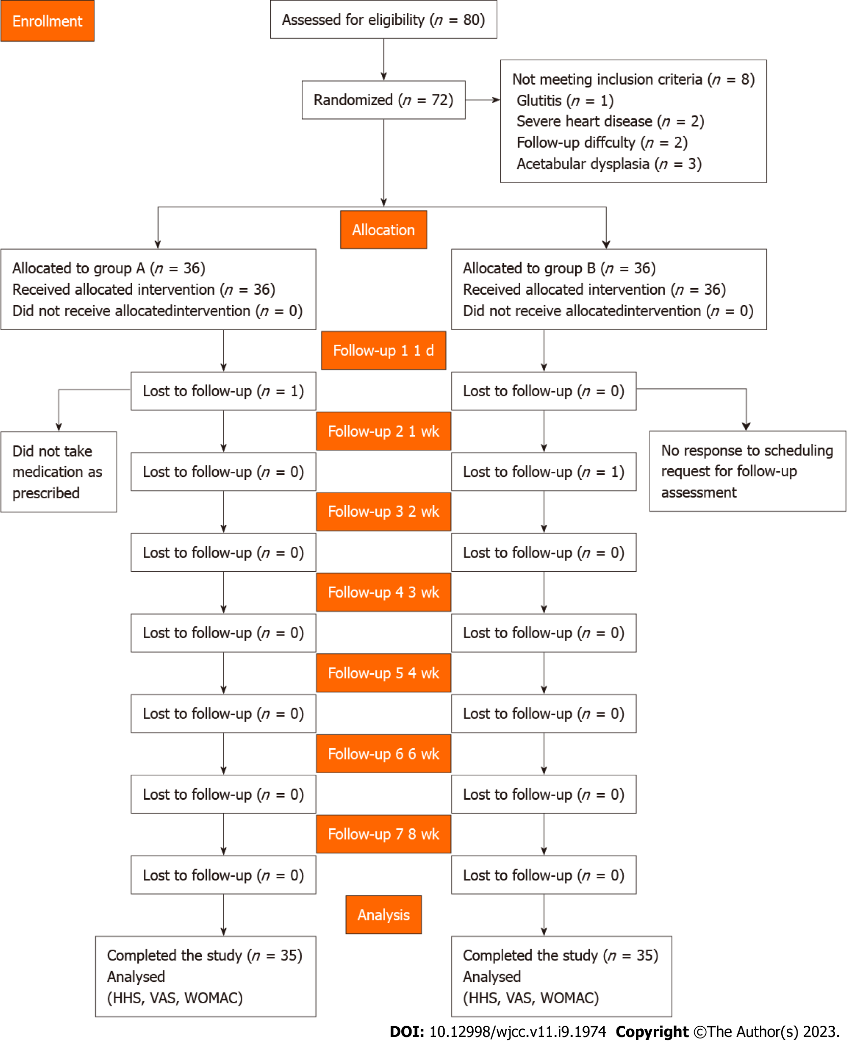
Figure 2 Study flow diagram.
HHS: Harris hip score; VAS: Visual analog scale; WOMAC: Western Ontario and McMaster Universities Osteoarthritis Index.
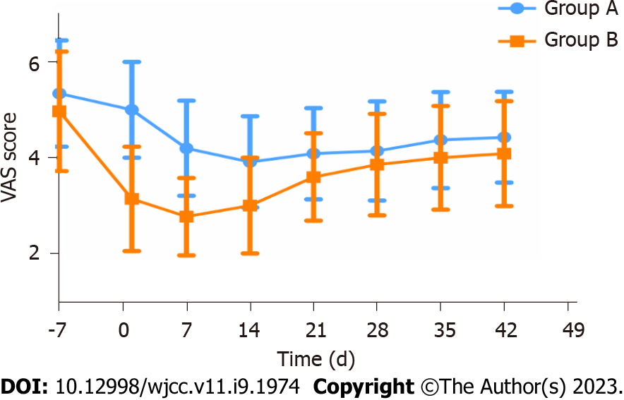
Figure 3 Changing trend of the visual analog scale in groups A and B.
VAS: Visual analog scale.
- Citation: Zhu JY, Yan J, Xiao J, Jia HG, Liang HJ, Xing GY. Effects of individual shock wave therapy vs celecoxib on hip pain caused by femoral head necrosis. World J Clin Cases 2023; 11(9): 1974-1984
- URL: https://www.wjgnet.com/2307-8960/full/v11/i9/1974.htm
- DOI: https://dx.doi.org/10.12998/wjcc.v11.i9.1974


