Copyright
©The Author(s) 2023.
World J Clin Cases. Feb 6, 2023; 11(4): 972-978
Published online Feb 6, 2023. doi: 10.12998/wjcc.v11.i4.972
Published online Feb 6, 2023. doi: 10.12998/wjcc.v11.i4.972
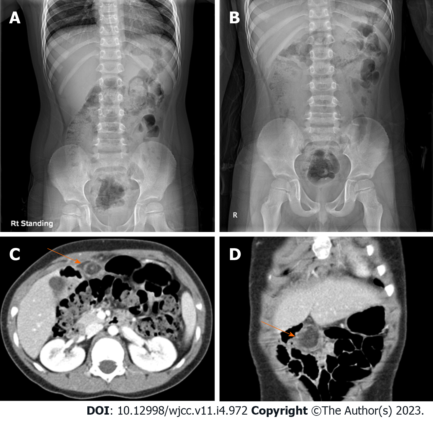
Figure 1 Radiologic study.
A and B: Simple abdomen; C and D: Computed tomography. On erect (A) and supine (B) plain radiography, feces and gas are found inside the large bowel, and no other specific findings are shown. Axial (C) and coronal (D) scan of abdominal computed tomography show a fat lobule below the umbilical ligament of the liver left lobe. A hyperdense halo and surrounding fat stranding are in the periphery of the fat lobule (arrow). The vessel of the upper portion inside the fat lobule shows a whirling sign.
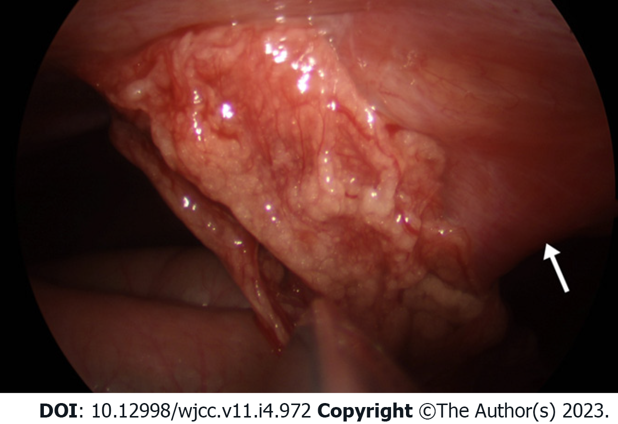
Figure 2 Intraoperative photography demonstrating an infarcted omentum adherent to the anterior abdominal wall.
The white arrow shows the falciform ligament.
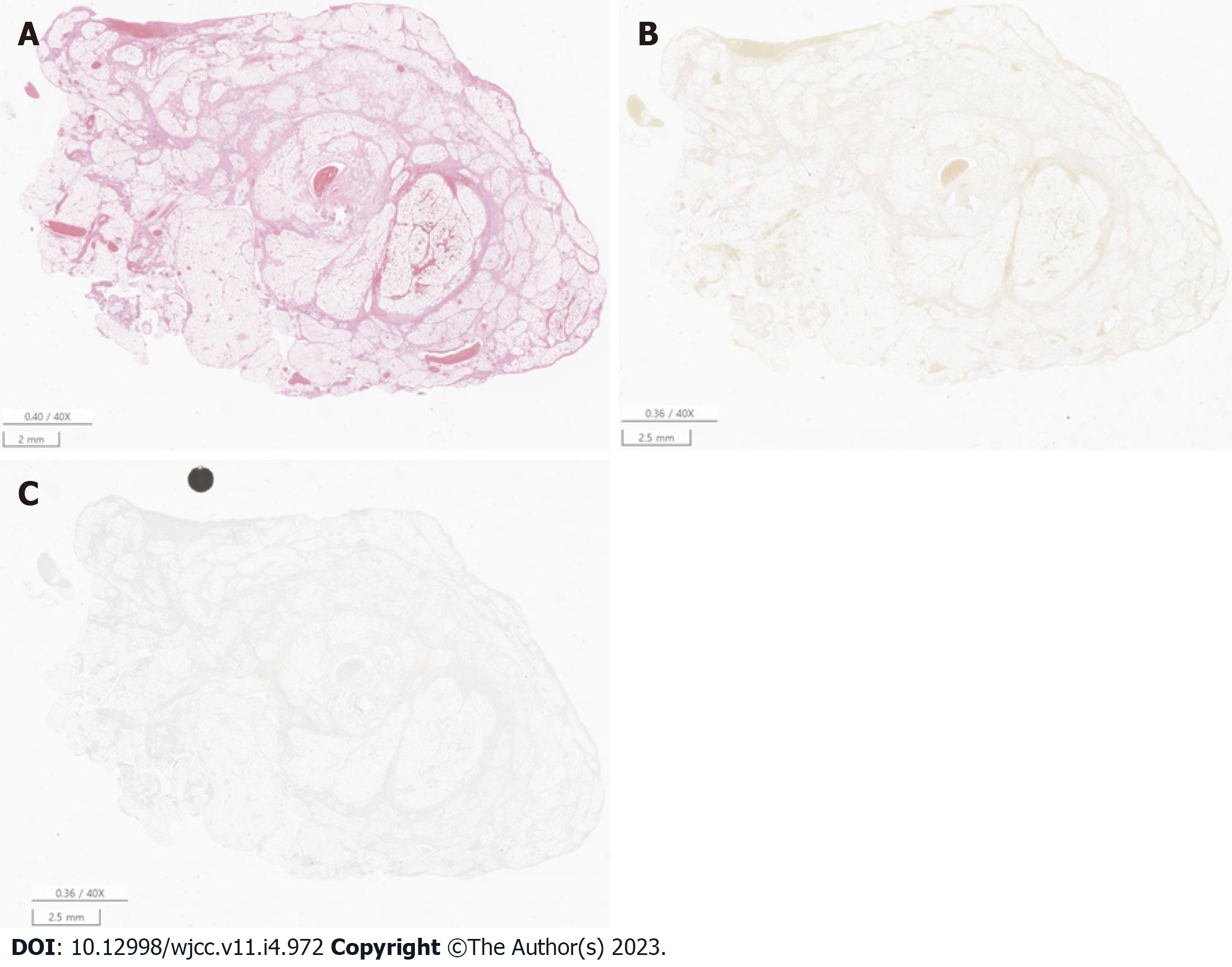
Figure 3 Representative microscopic features.
A: Omentum is consistent with a hemorrhagic infarction [Hematoxylin & Eosin staining (HE), original magnification × 40]; B: Microorganisms were not identified by Gram staining (HE, original magnification × 40); C: The Ki-67 proliferative index was 5% (HE, original magnification × 40).
- Citation: Hwang JK, Cho YJ, Kang BS, Min KW, Cho YS, Kim YJ, Lee KS. Omental infarction diagnosed by computed tomography, missed with ultrasonography: A case report. World J Clin Cases 2023; 11(4): 972-978
- URL: https://www.wjgnet.com/2307-8960/full/v11/i4/972.htm
- DOI: https://dx.doi.org/10.12998/wjcc.v11.i4.972


