Copyright
©The Author(s) 2023.
World J Clin Cases. Feb 6, 2023; 11(4): 945-951
Published online Feb 6, 2023. doi: 10.12998/wjcc.v11.i4.945
Published online Feb 6, 2023. doi: 10.12998/wjcc.v11.i4.945
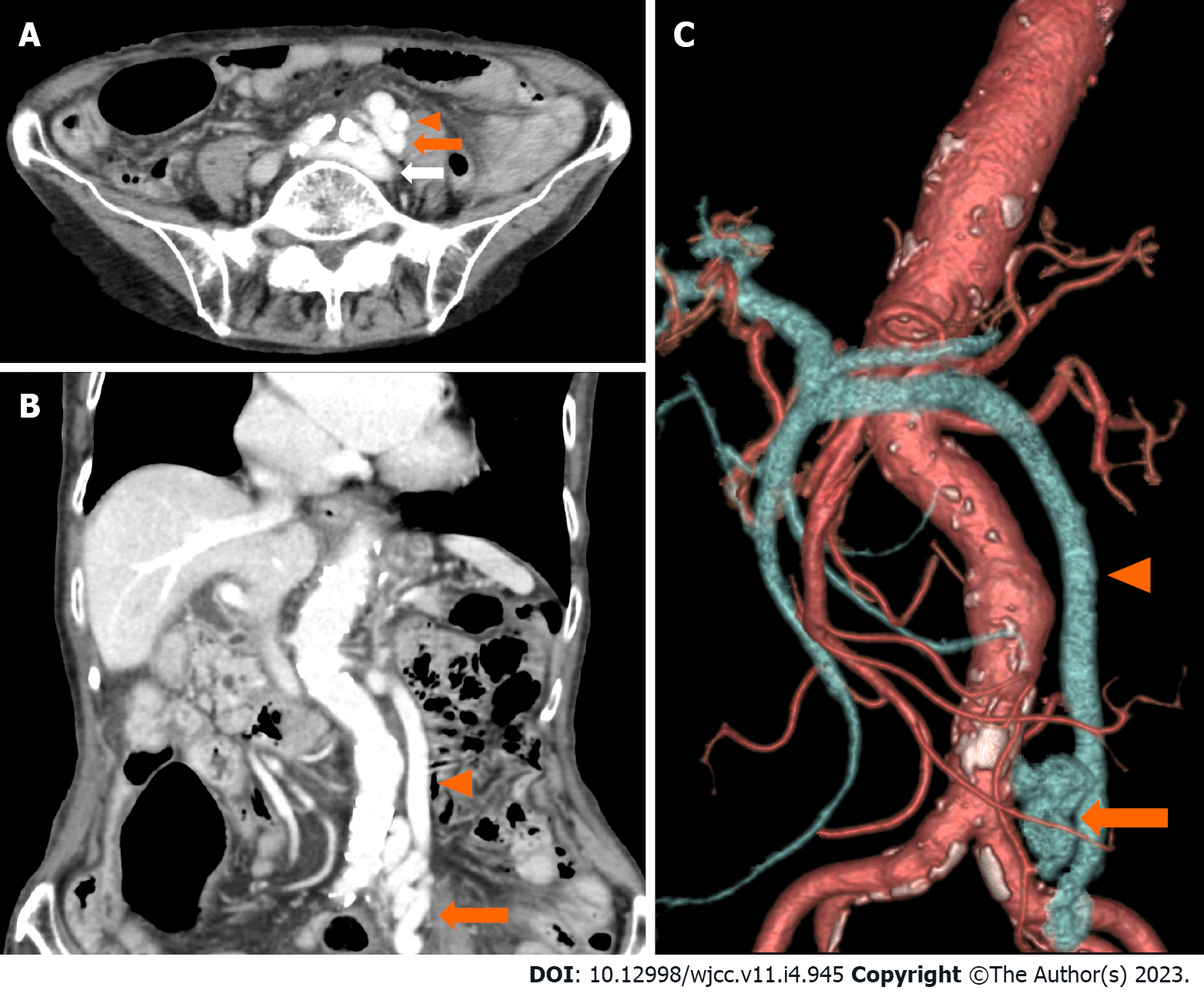
Figure 1 Abdominal computed tomography scan with contrast enhancement.
A-C: Images showing a portosystemic shunt (orange arrow) running between the left common iliac vein (white arrow) and the inferior mesenteric vein (arrowhead).
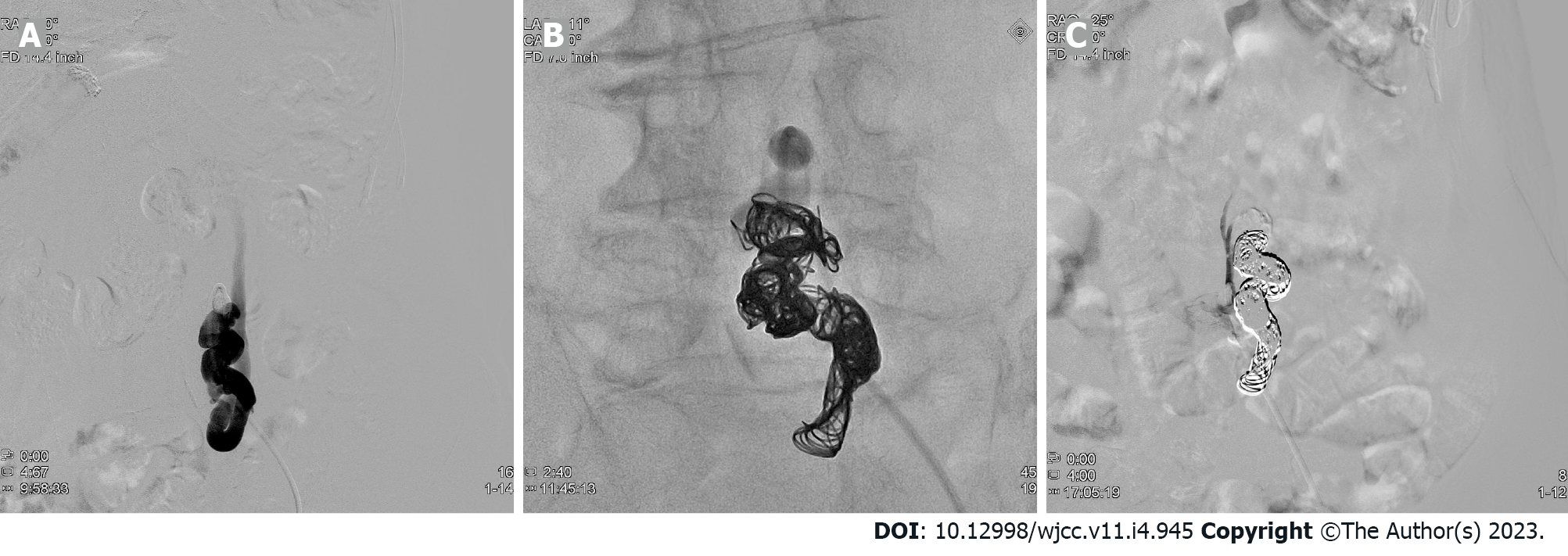
Figure 2 Angiography images before and after embolization.
A: A balloon catheter is advanced into the portosystemic shunt through the left femoral vein; B: The shunt is filled with the coil; C: The shunt is completely obliterated.
- Citation: Nishi A, Kenzaka T, Sogi M, Nakaminato S, Suzuki T. Treatment of portosystemic shunt-borne hepatic encephalopathy in a 97-year-old woman using balloon-occluded retrograde transvenous obliteration: A case report. World J Clin Cases 2023; 11(4): 945-951
- URL: https://www.wjgnet.com/2307-8960/full/v11/i4/945.htm
- DOI: https://dx.doi.org/10.12998/wjcc.v11.i4.945


