Copyright
©The Author(s) 2023.
World J Clin Cases. Feb 6, 2023; 11(4): 938-944
Published online Feb 6, 2023. doi: 10.12998/wjcc.v11.i4.938
Published online Feb 6, 2023. doi: 10.12998/wjcc.v11.i4.938
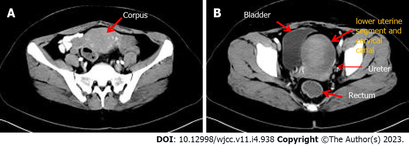
Figure 1 Computerized tomography imaging.
A: Uterine body; B: Lower uterine segment and cervical canal lesion.
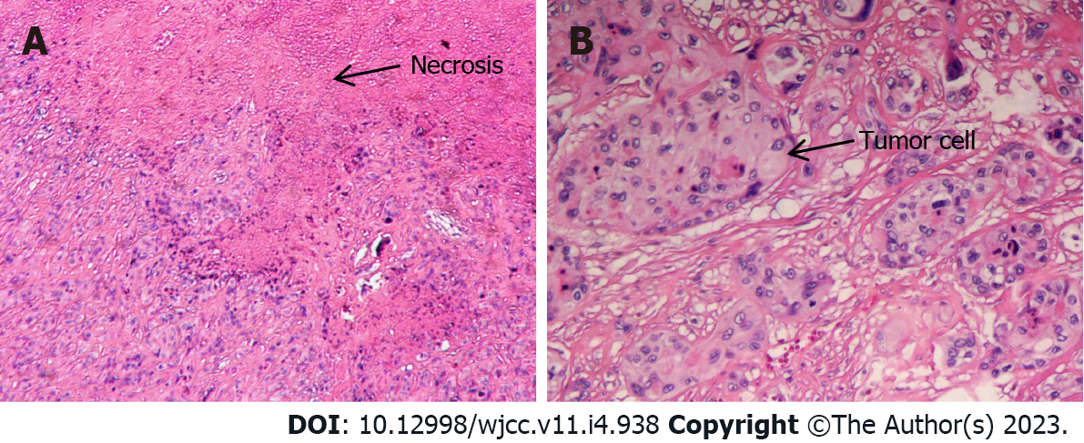
Figure 2 Histological analysis: Hematoxylin-eosin stain.
A: Geographical necrosis of tumors (×40); B: Mononuclear intermediate trophoblast cells with vacuoles and prominent nucleoli (×100).
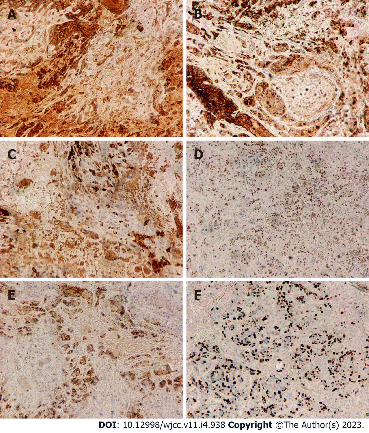
Figure 3 Immunohistochemistry assay (×40).
A: Human chorionic gonadotropin -positive; B: Epithelial membrane antigen -positive; C: Inhibin-alpha-positive; D: p63-positive; E: Placental alkaline phosphatase -positive; F: Ki-67 proliferation index of 40%.
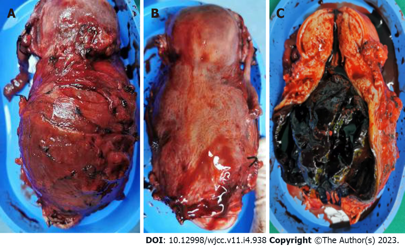
Figure 4 Surgical specimen.
A: Front view; B: Rear view; C: Anterior wall section.
- Citation: Yuan LQ, Hao T, Pan GY, Guo H, Li DP, Liu NF. Epithelioid trophoblastic tumor of the lower uterine segment and cervical canal: A case report. World J Clin Cases 2023; 11(4): 938-944
- URL: https://www.wjgnet.com/2307-8960/full/v11/i4/938.htm
- DOI: https://dx.doi.org/10.12998/wjcc.v11.i4.938


