Copyright
©The Author(s) 2023.
World J Clin Cases. Feb 6, 2023; 11(4): 909-917
Published online Feb 6, 2023. doi: 10.12998/wjcc.v11.i4.909
Published online Feb 6, 2023. doi: 10.12998/wjcc.v11.i4.909
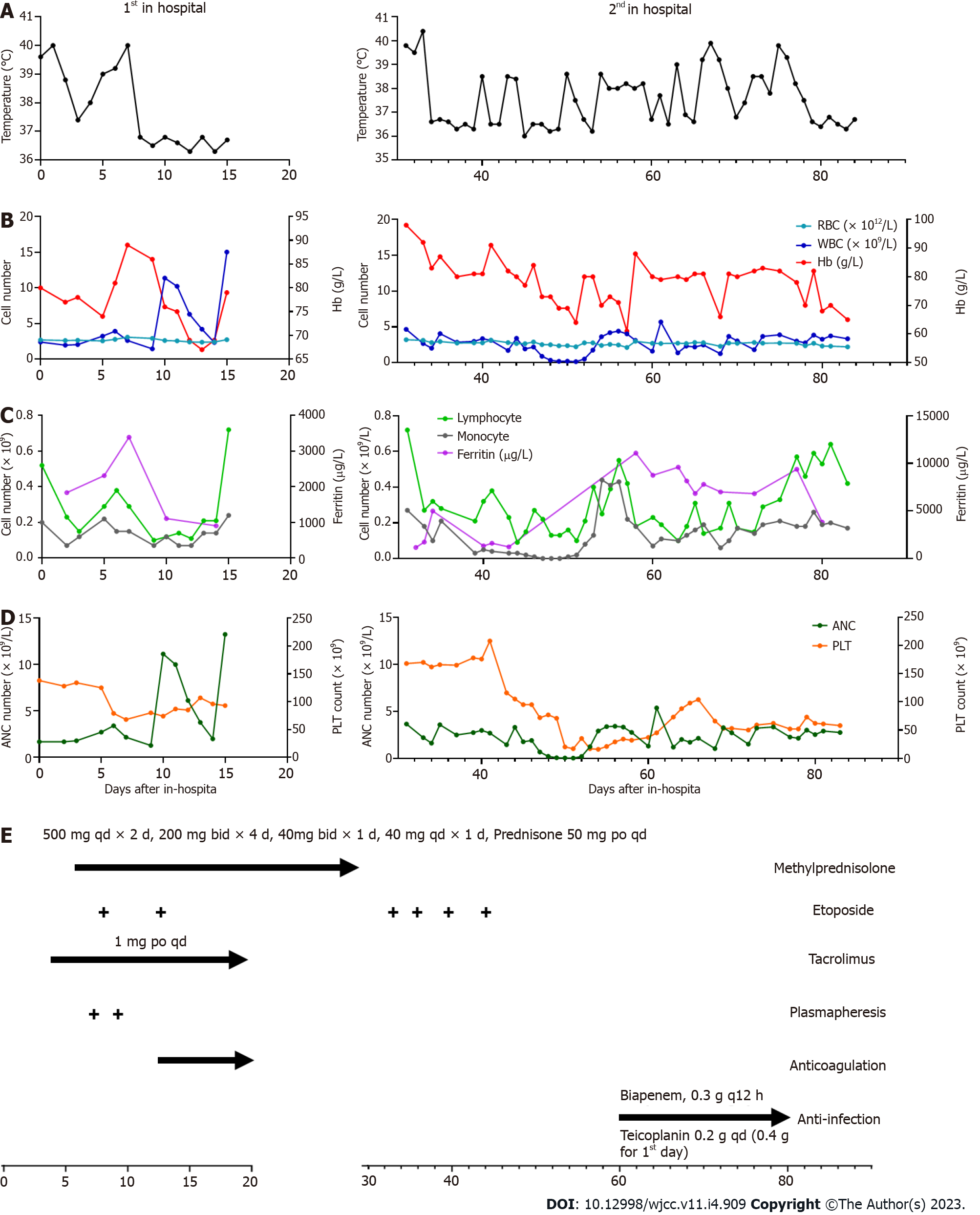
Figure 1 Inflammatory and hematologic results during hospitalization.
A: Body temperature of the patients; B-C: the count of blood cells, including red blood cells (RBC), white blood cells (WBC), lymphocyte and monocyte; D: The concentration of hemoglobin and ferritin; E: Treatment schedule, lines and arrows represent continuous therapy. RBC: Red blood cell; WBC: White blood cell; Hb: Hemoglobin; PLT: Platelet; ANC: Absolute neutrophilic count.
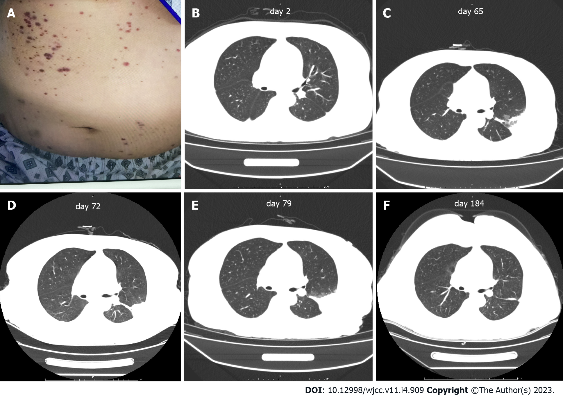
Figure 2 Abdominal skin and scan of chest computed tomography.
A: Scattered petechiae and ecchymoses were detected over the patient’s abdomen and extremities during hospitalization; B-F: Chest computed tomography showed the recovery of infection foci in the lungs.
- Citation: Peng LY, Liu JB, Zuo HJ, Shen GF. Unusual presentation of systemic lupus erythematosus as hemophagocytic lymphohistiocytosis in a female patient: A case report. World J Clin Cases 2023; 11(4): 909-917
- URL: https://www.wjgnet.com/2307-8960/full/v11/i4/909.htm
- DOI: https://dx.doi.org/10.12998/wjcc.v11.i4.909


