Copyright
©The Author(s) 2023.
World J Clin Cases. Feb 6, 2023; 11(4): 844-851
Published online Feb 6, 2023. doi: 10.12998/wjcc.v11.i4.844
Published online Feb 6, 2023. doi: 10.12998/wjcc.v11.i4.844
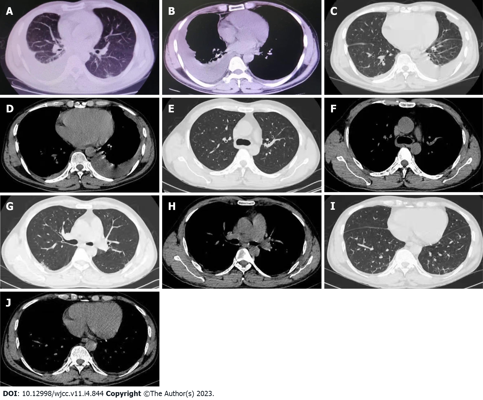
Figure 1 Chest computed tomography image.
A and B: Chest computed tomography (CT) scan performed on May 14; C and D: Chest CT scan performed on May 26; E-J: Chest CT scan performed on June 22.
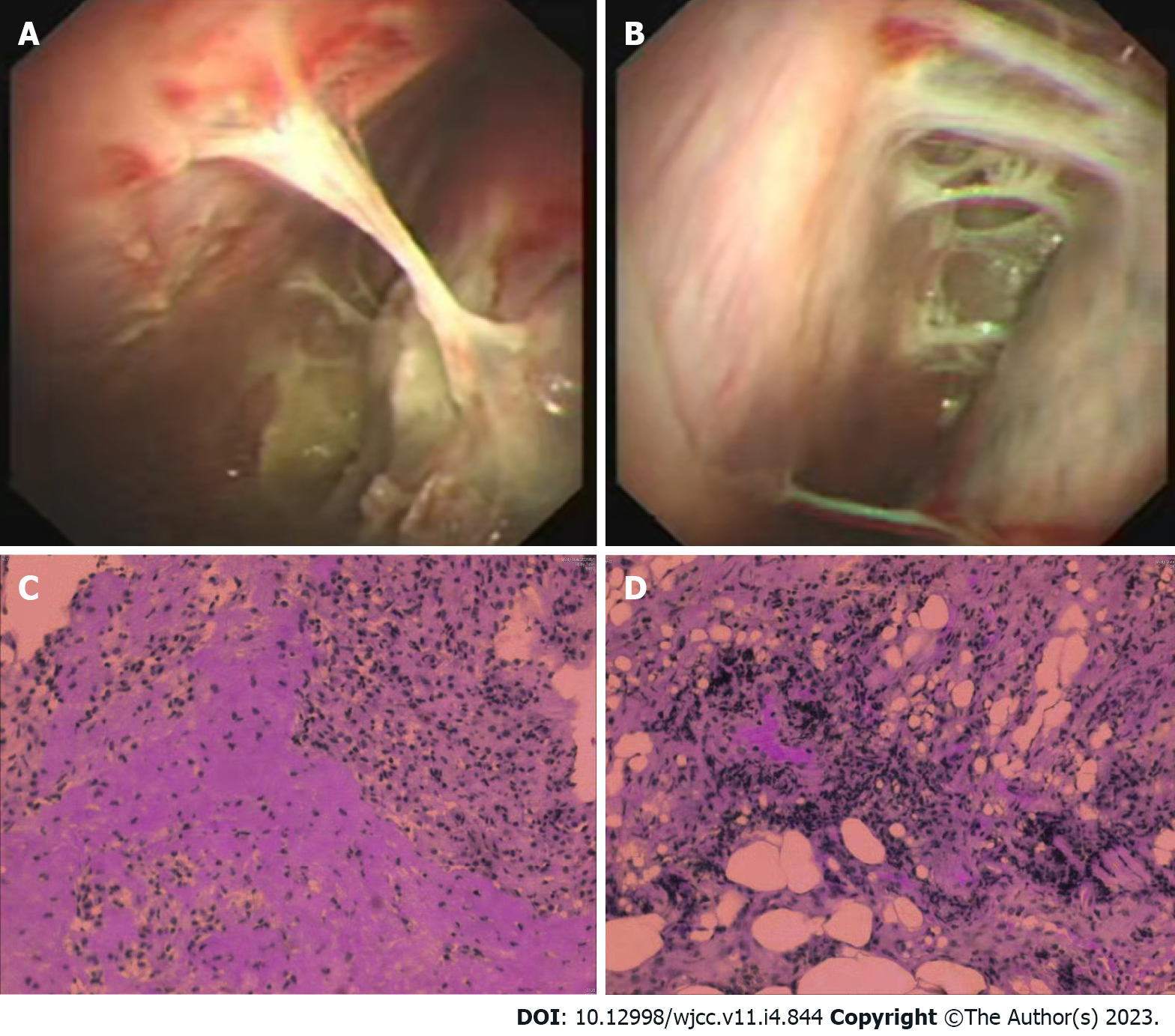
Figure 2 Thoracoscopy and pathologic image.
A and B: Thoracoscopy image performed on May 28; C and D: Pathologic findings of the pleural biopsy on May 30.
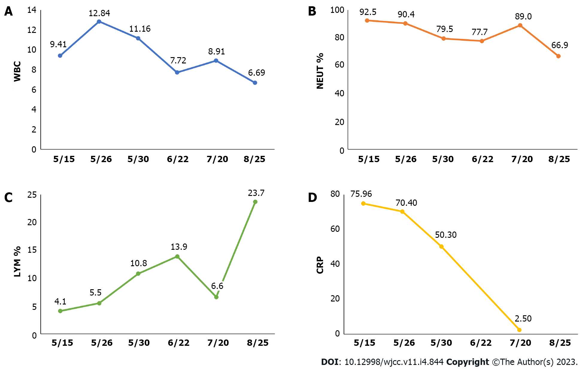
Figure 3 Course of some laboratory results of this patient over time.
A: The course of white blood cell over time; B: The course of neutrophils % over time; C: The course of lymphocytes % over time; D: The course of C-reactive protein over time. WBC: White blood cell; NEUT: Neutrophils; LYM: Lymphocytes; CRP: C-reactive protein.
- Citation: Liu XP, Mao CX, Wang GS, Zhang MZ. Metagenomic next-generation sequencing for pleural effusions induced by viral pleurisy: A case report. World J Clin Cases 2023; 11(4): 844-851
- URL: https://www.wjgnet.com/2307-8960/full/v11/i4/844.htm
- DOI: https://dx.doi.org/10.12998/wjcc.v11.i4.844


